Pap smear. HSIL. (Papanicolaou x100)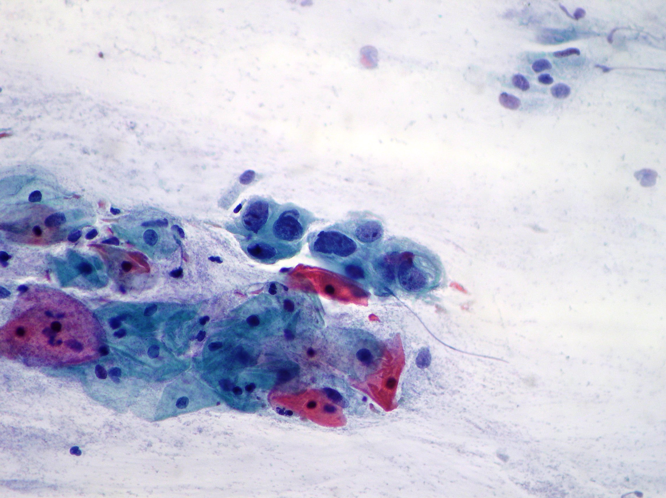
Pap smear. HSIL with involvment of endocervical channel. (Papanicolaou x200)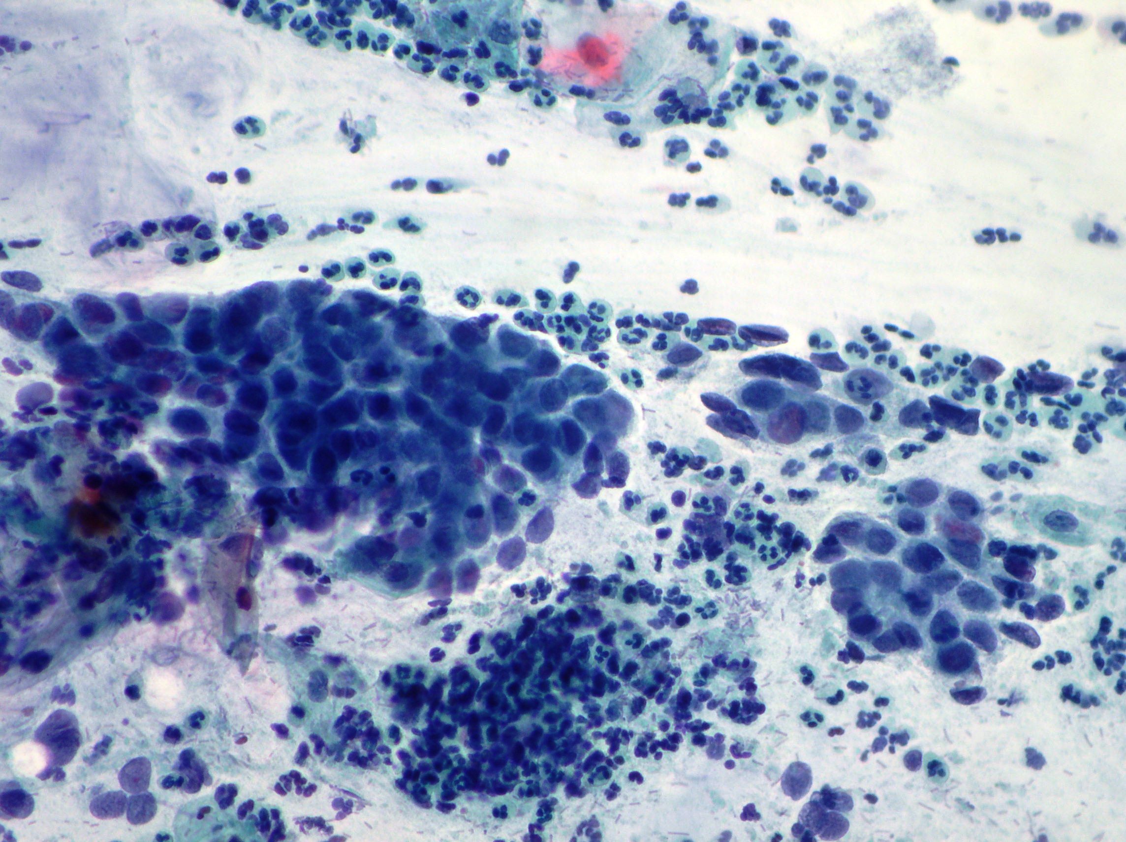
Pap smear. ASC-US. (Papanicolaou x200)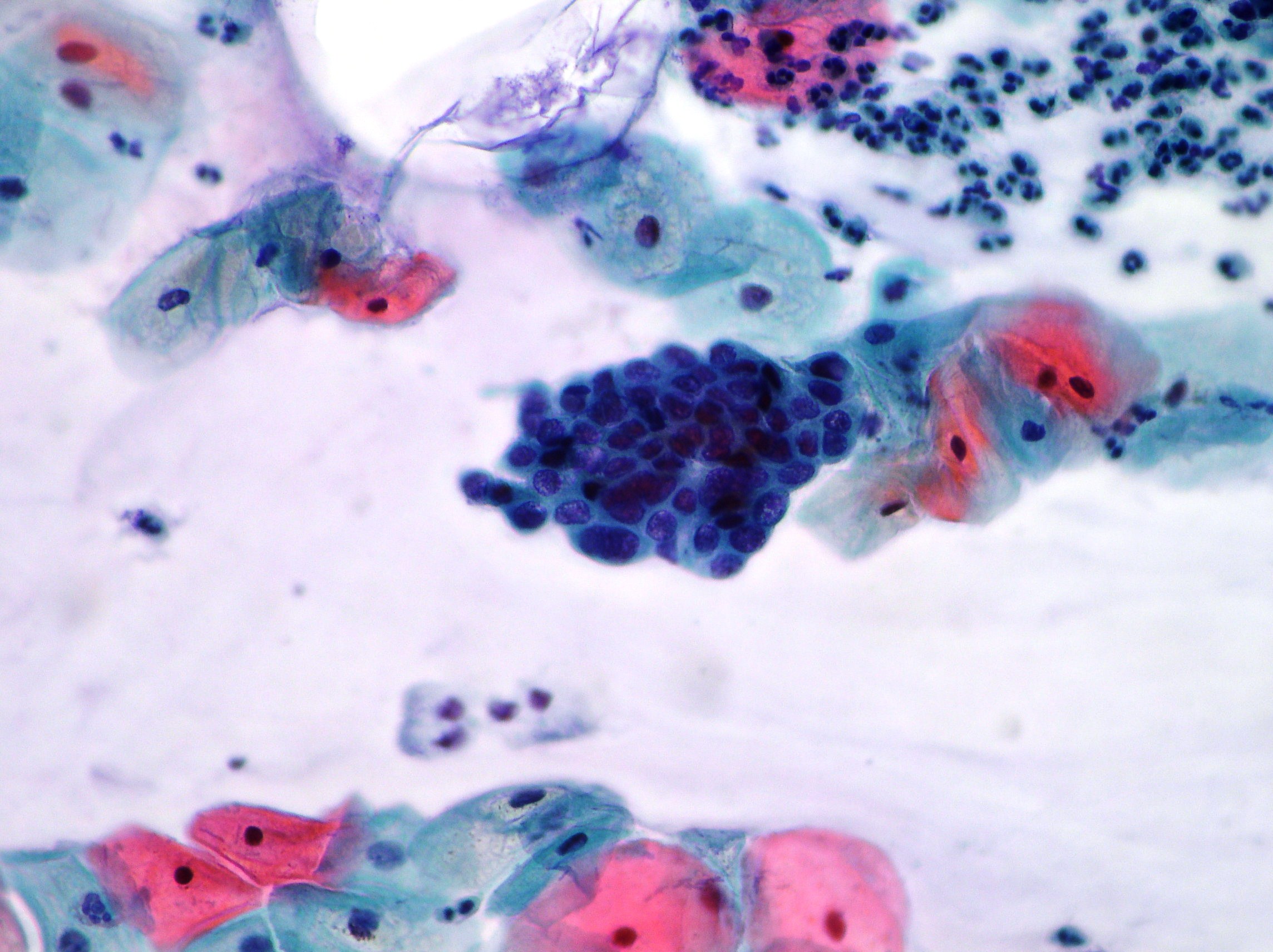
Pap smear. Mature and immature squamous metaplasia showing cells organized singly and on a sheet with relatively large nuclei and vacuolated cytoplasms. (Papanicolaou, x200)
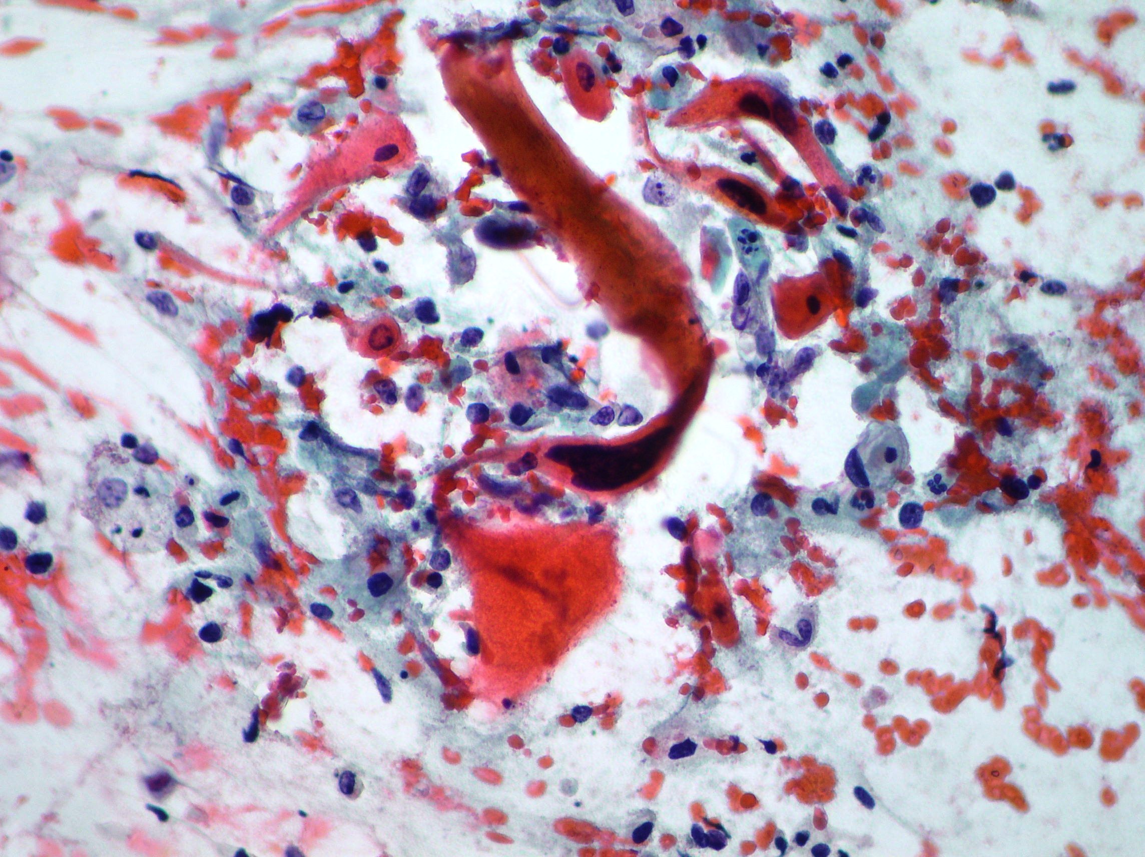
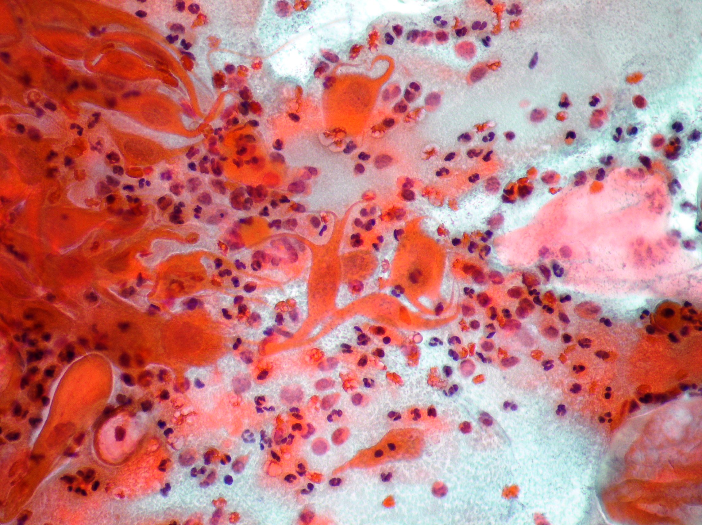
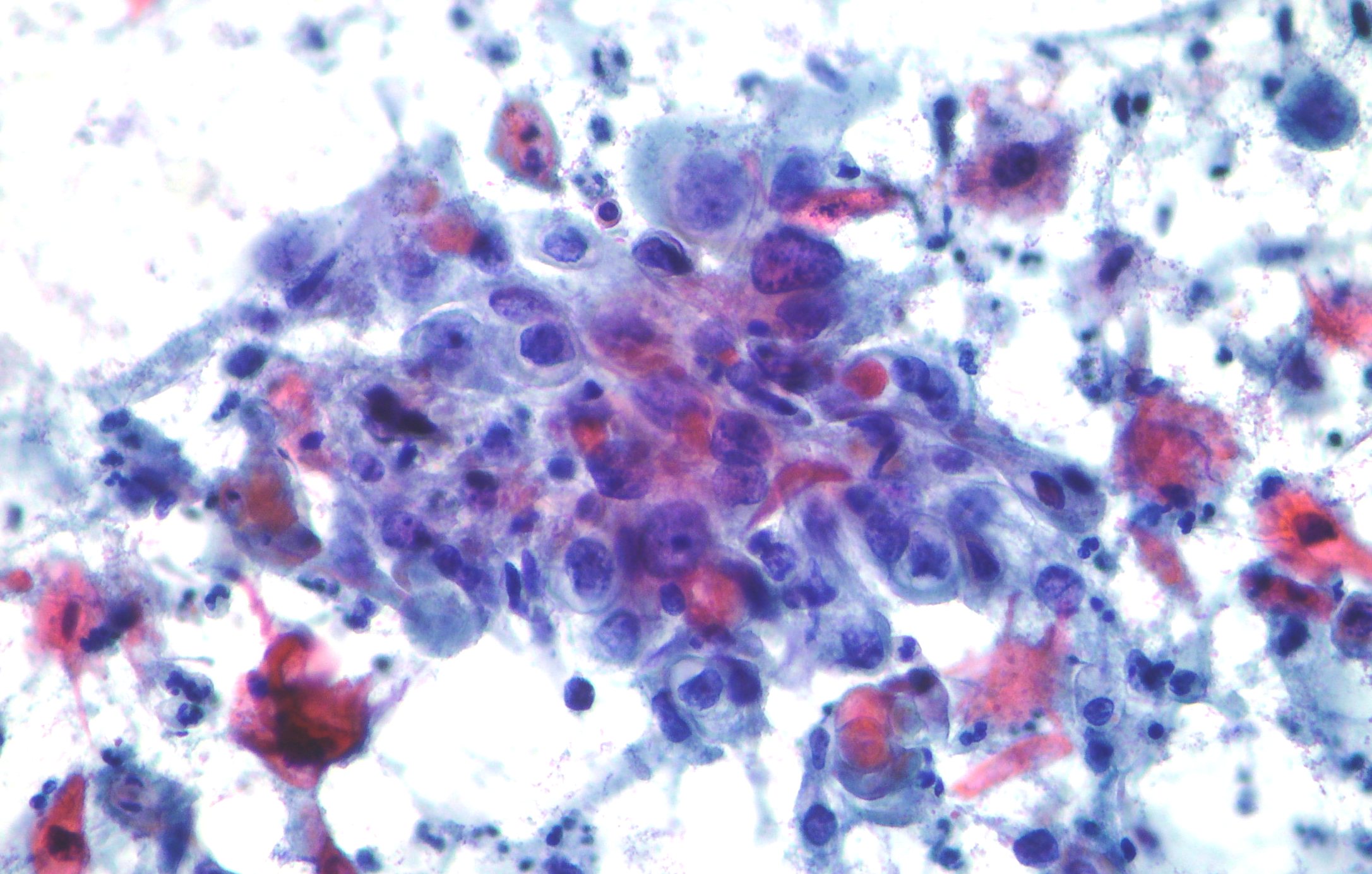
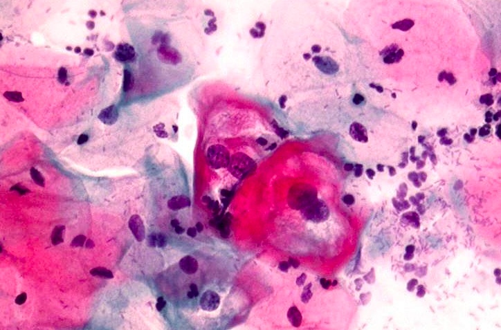
Pap smear. Keratinizing squamous cell carcinoma showing bizzarre shaped cytoplasms and hyperchromatic nuclei. (Papanicolaou, x200)
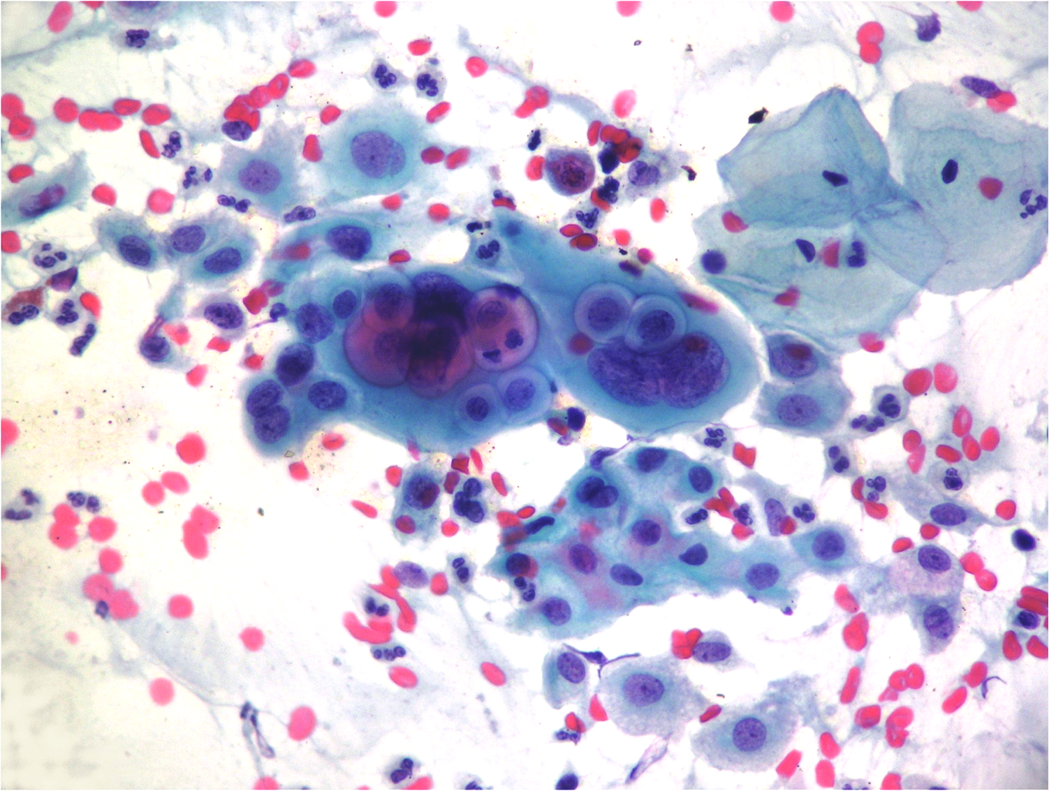
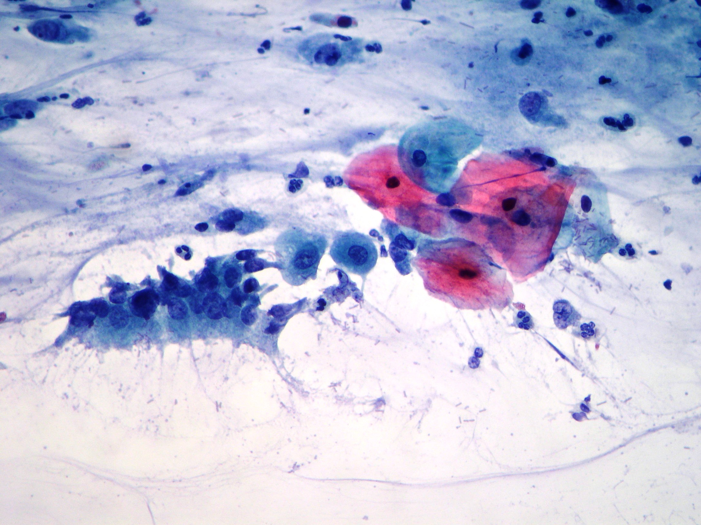
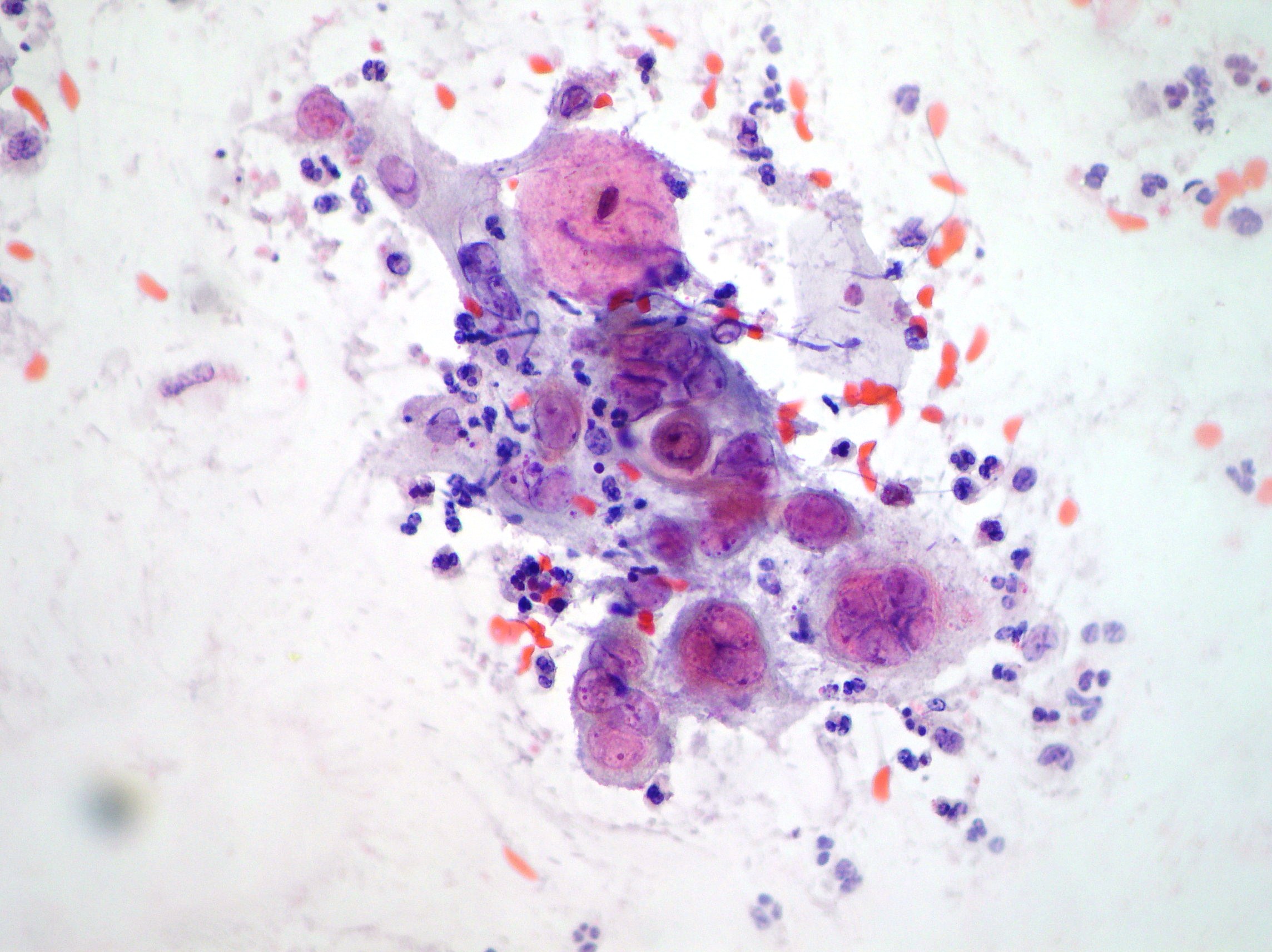
Pap smear. Radiation changes in the uterine cervix exhibiting enlarged multinucleate cells with bizzarre shapes, vacuolated cytoplasms and phagocitosis of neutrophils. (Papanicolaou, 200)
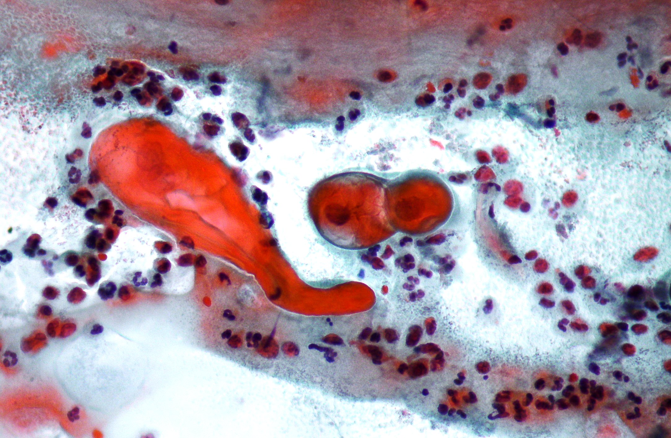
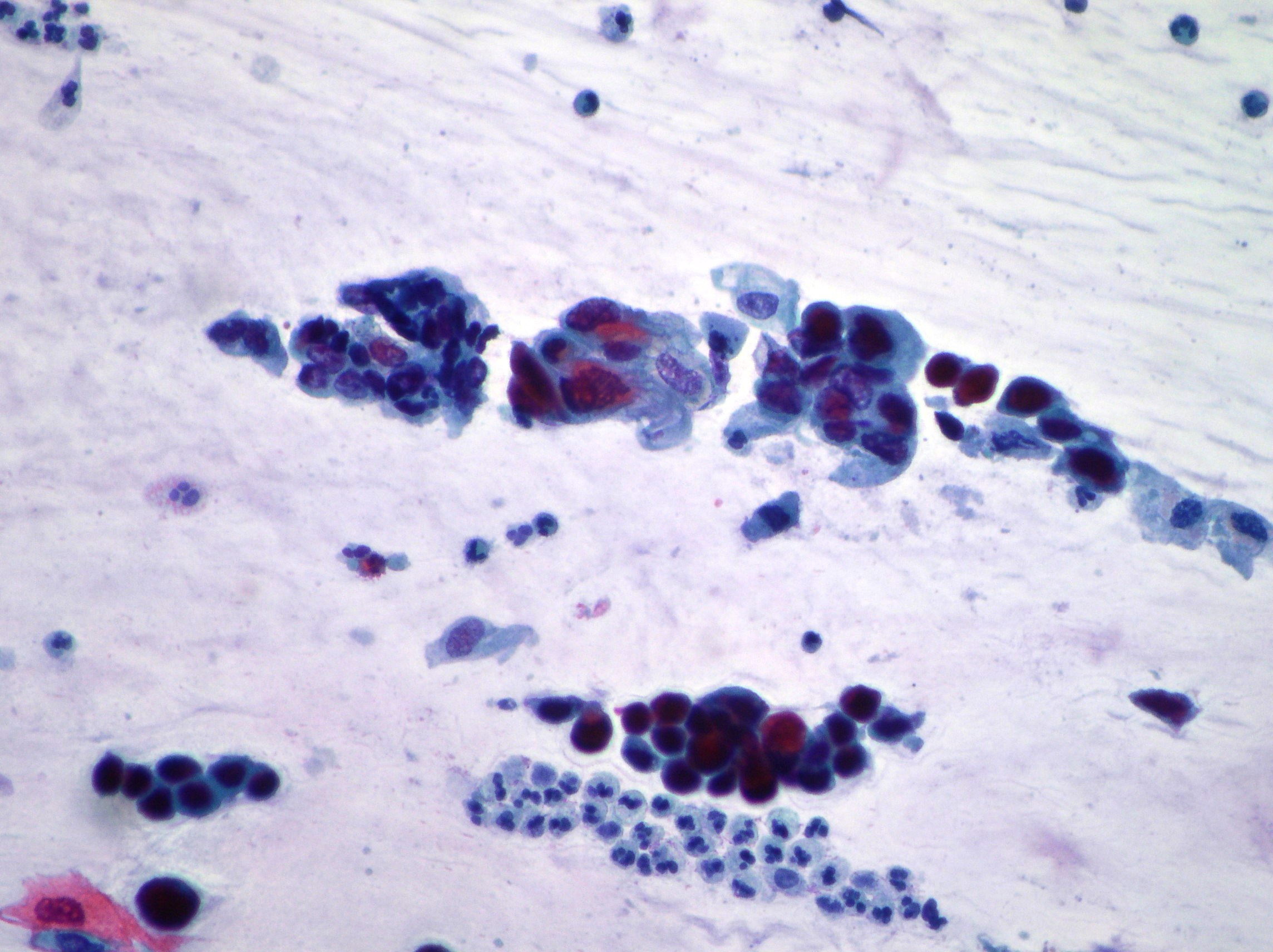
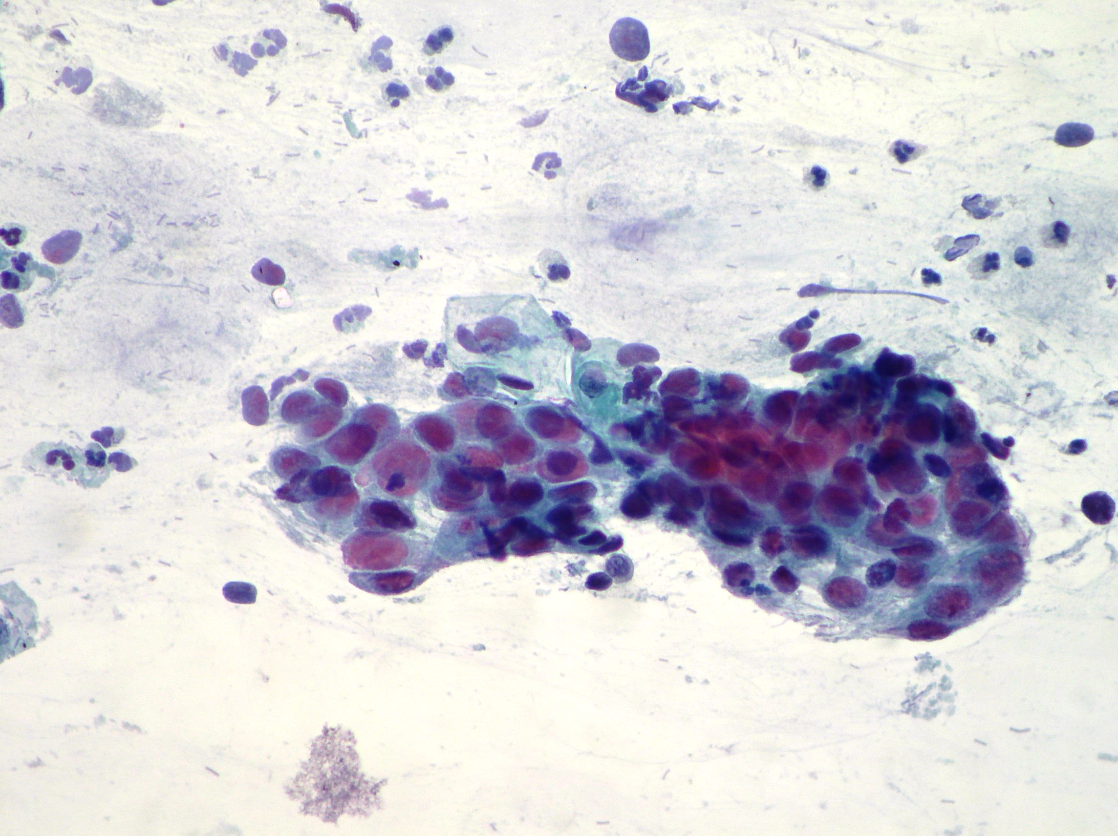
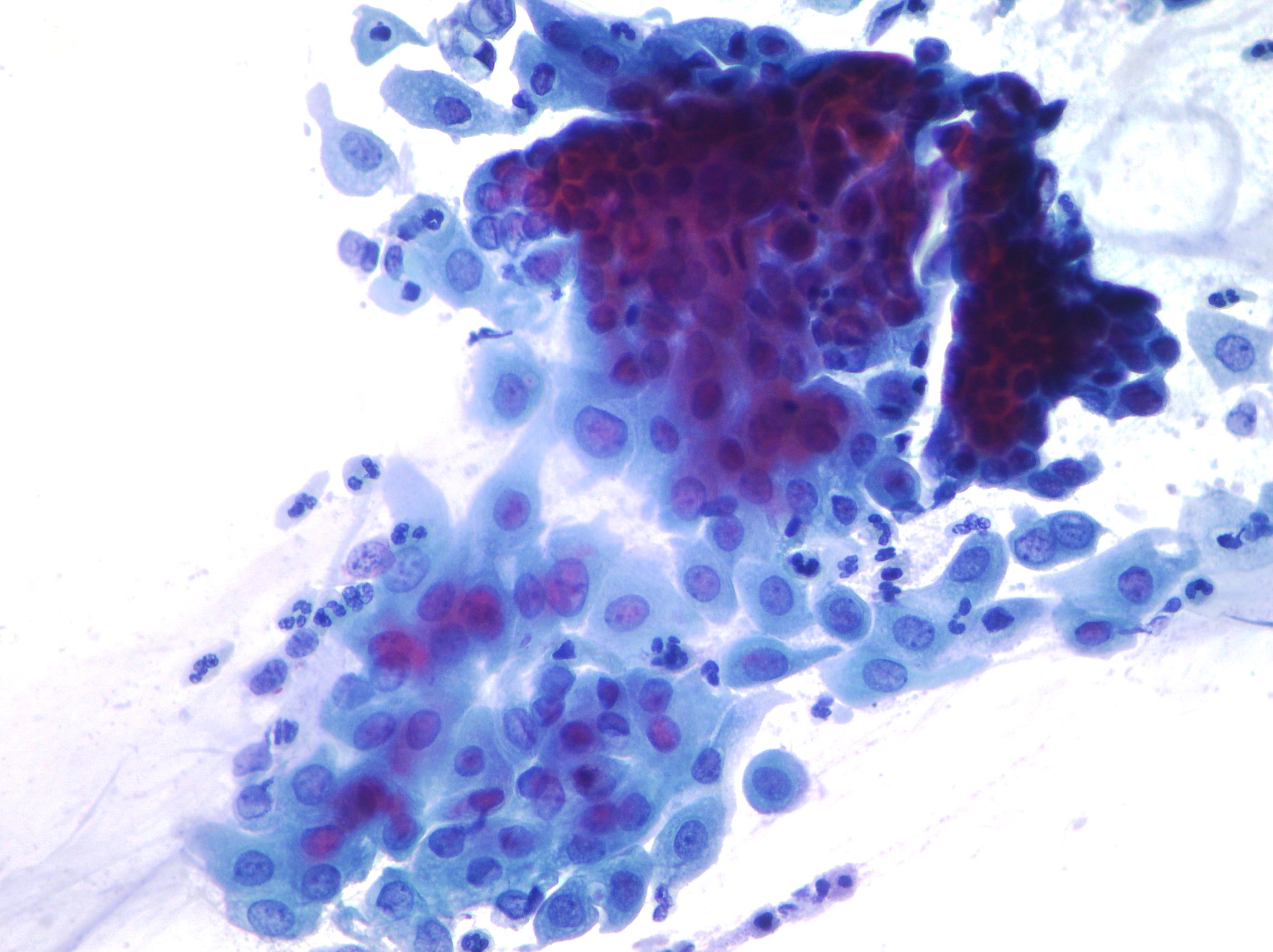
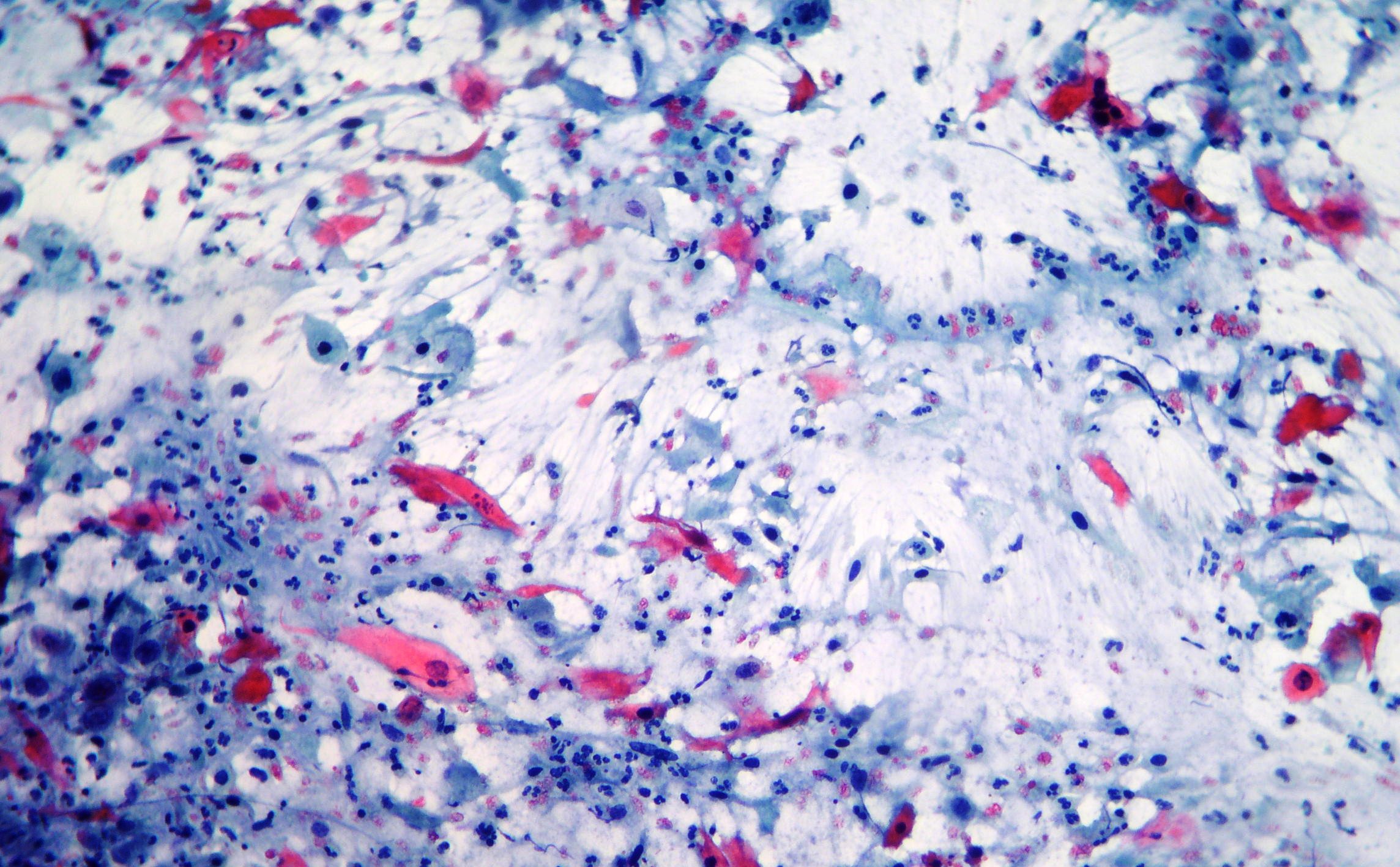
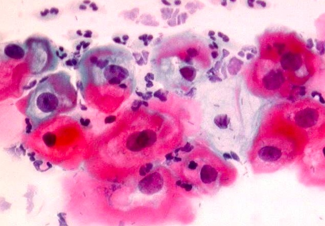
Pap smear. Keratinizing squamous cell carcinoma. Elongated and bizzarre shaped cells, degenerated round and oval nuclei with dense eosinophilic cytoplasm. Inflammation, debris and blood cells in the background. (Papanicolaou, x200, x100)
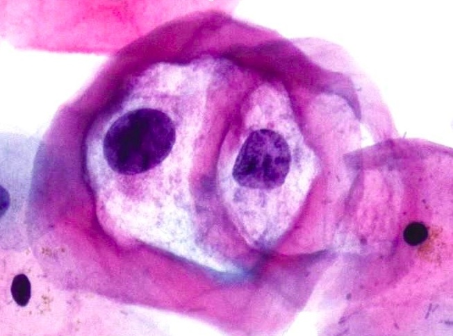
Pap smear. Typical koilocytotic atypia exhibiting large perinuclear halos, condensation of the chromatin and slight atypia. (Papanicolaou, x600)
Pap smear. Cervical smears with LSIL showing cells with vacuolization of the cytoplasm and slight atypia of the nuclei. (Papanicolaou, x400)
Pap smear. Normal superficial and intermediate squamous cell and tall columnar cells with eccentrically located nuclei. (Papanicolaou, x200)
Pap smear. Herpes genitalis infection (HSV) showing atypical multinucleation, molding and small intranuclear chromatin granules suggestive of an early infection. (Papanicolaou, x200)
Pap smear. Parabasal and basal cellsshowing hyperchromatic nucleus and high nucleocytoplasmic ratio suggestive of HSIL. (Papanicolaou, x200)