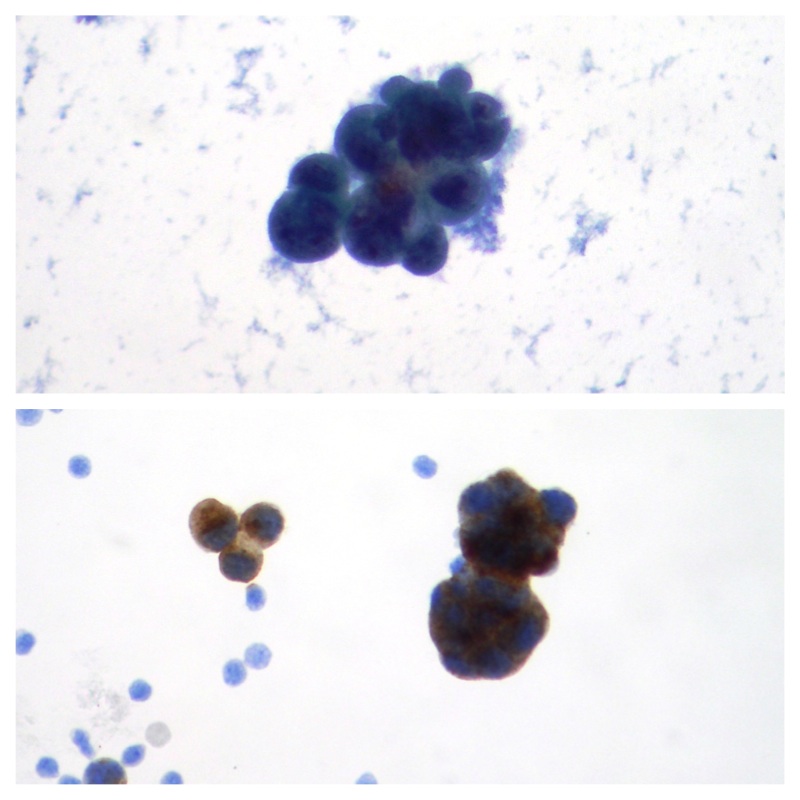
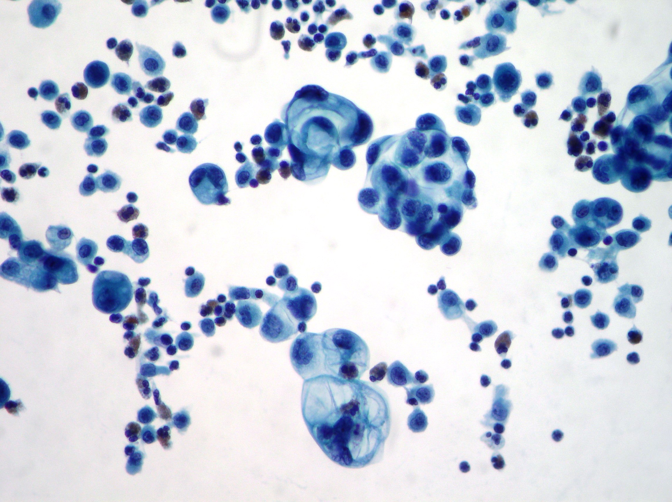
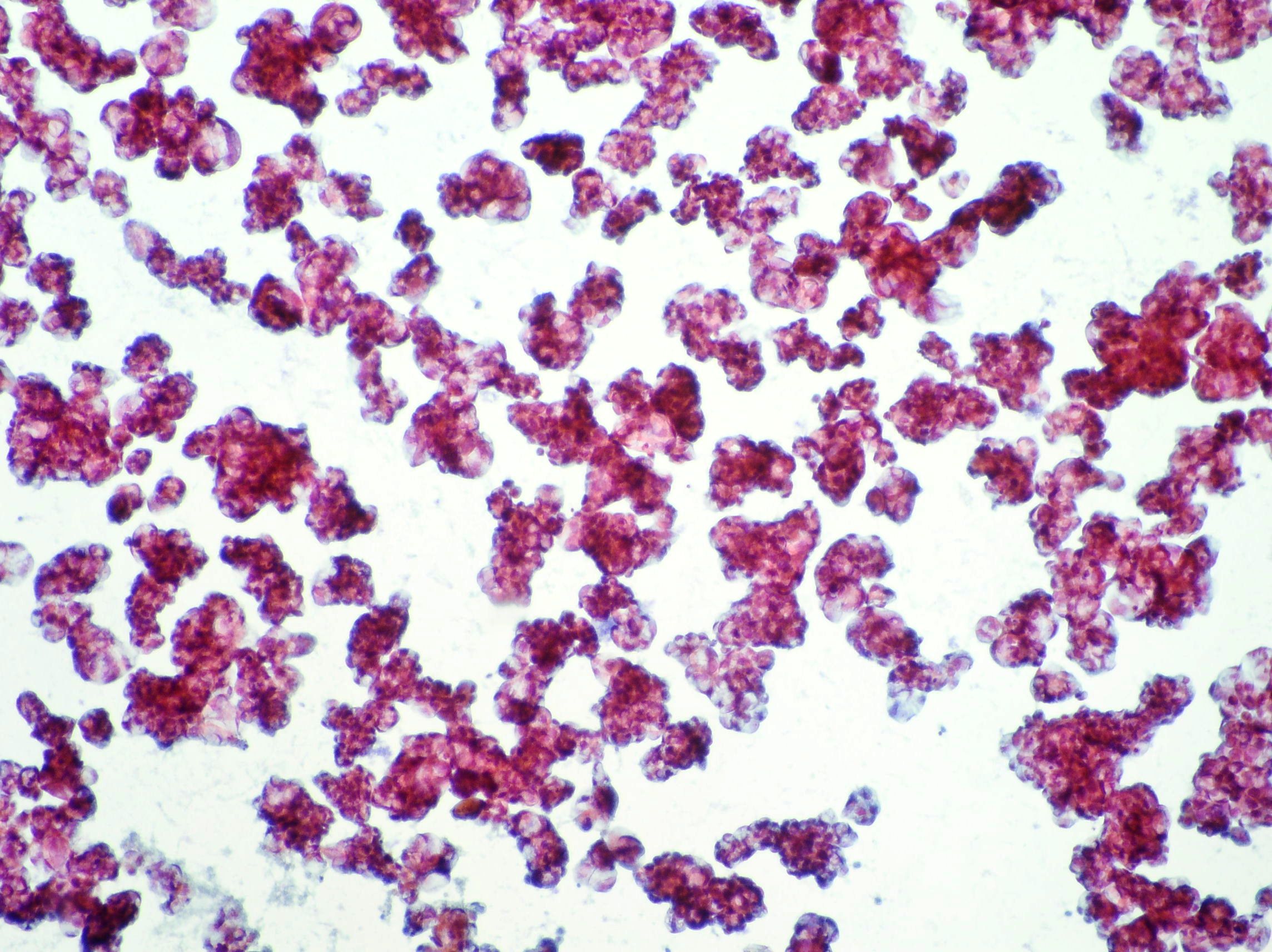
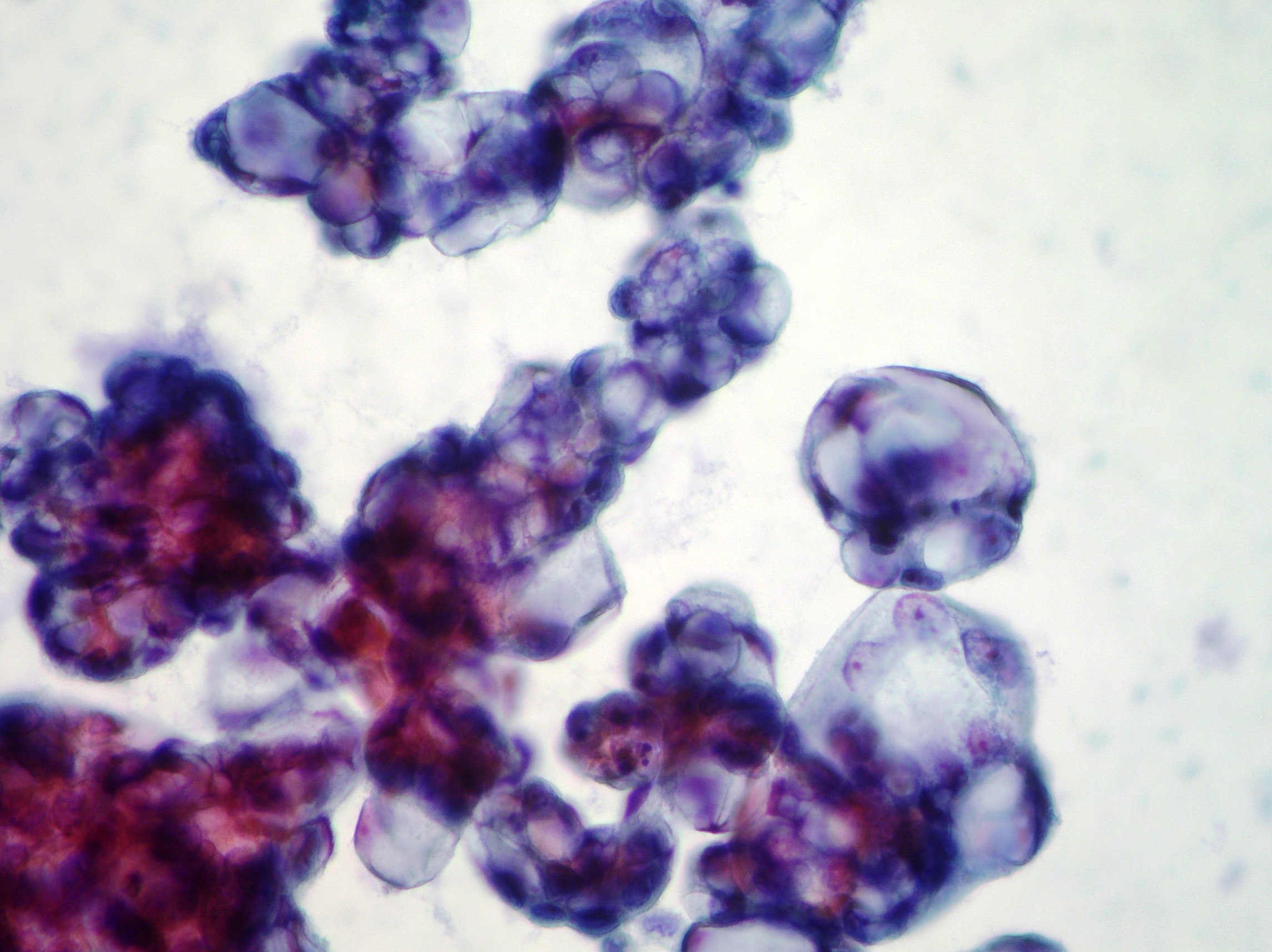
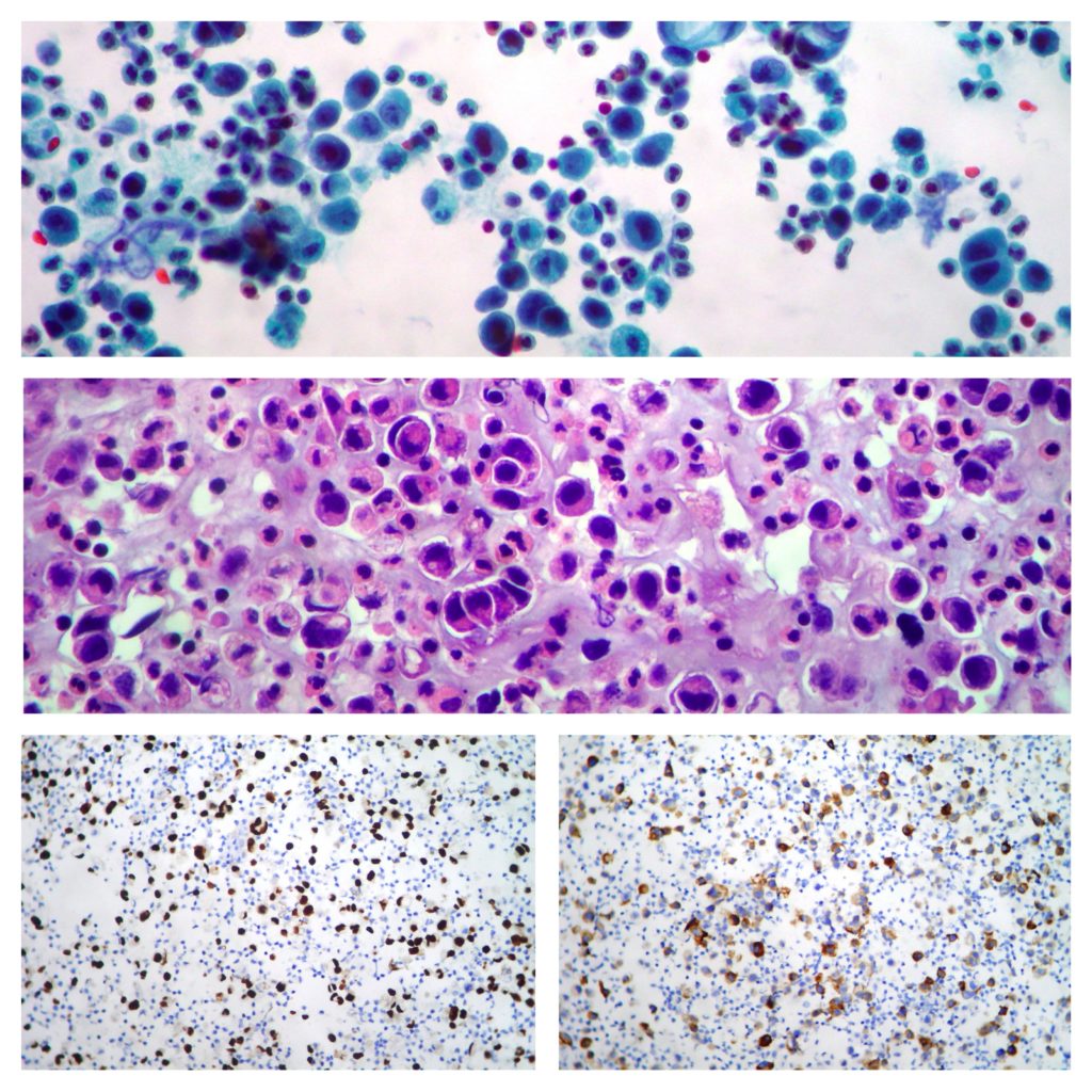
Metastatic urothelial carcinoma pleural effusion showing both GATA 3 (left side) and Uroplakin (right side) positive by immunohistochemistry. (Papanicolaou, H&E x200)
Pleural effusion
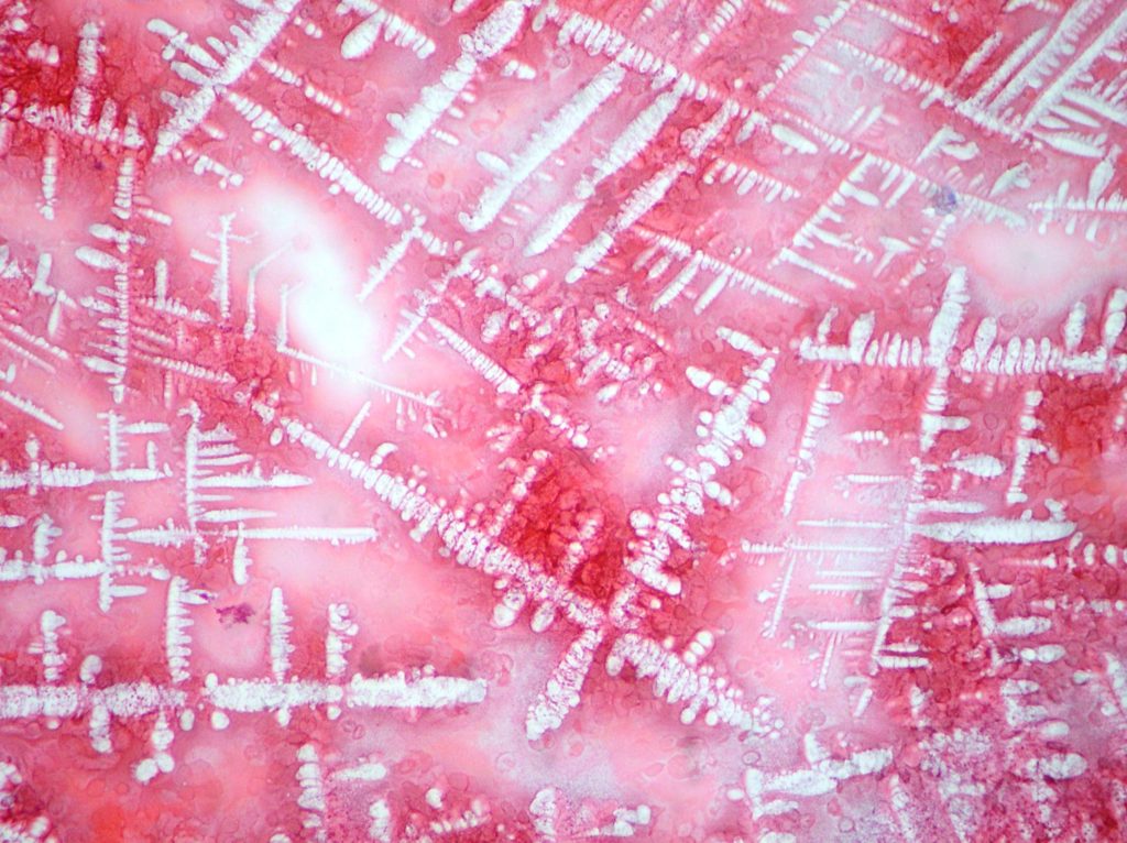
Artefacts of fixation/staining. (Papanicolaou x200)
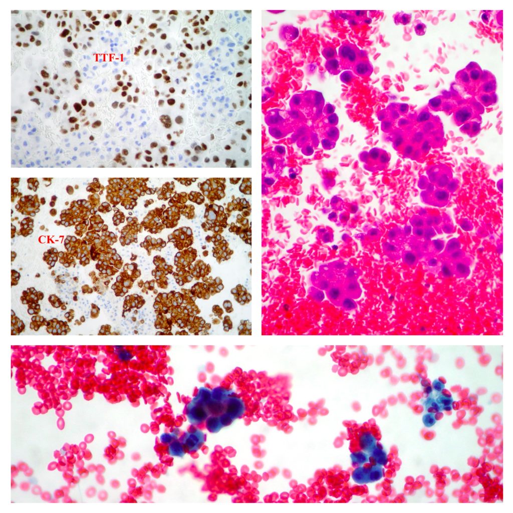
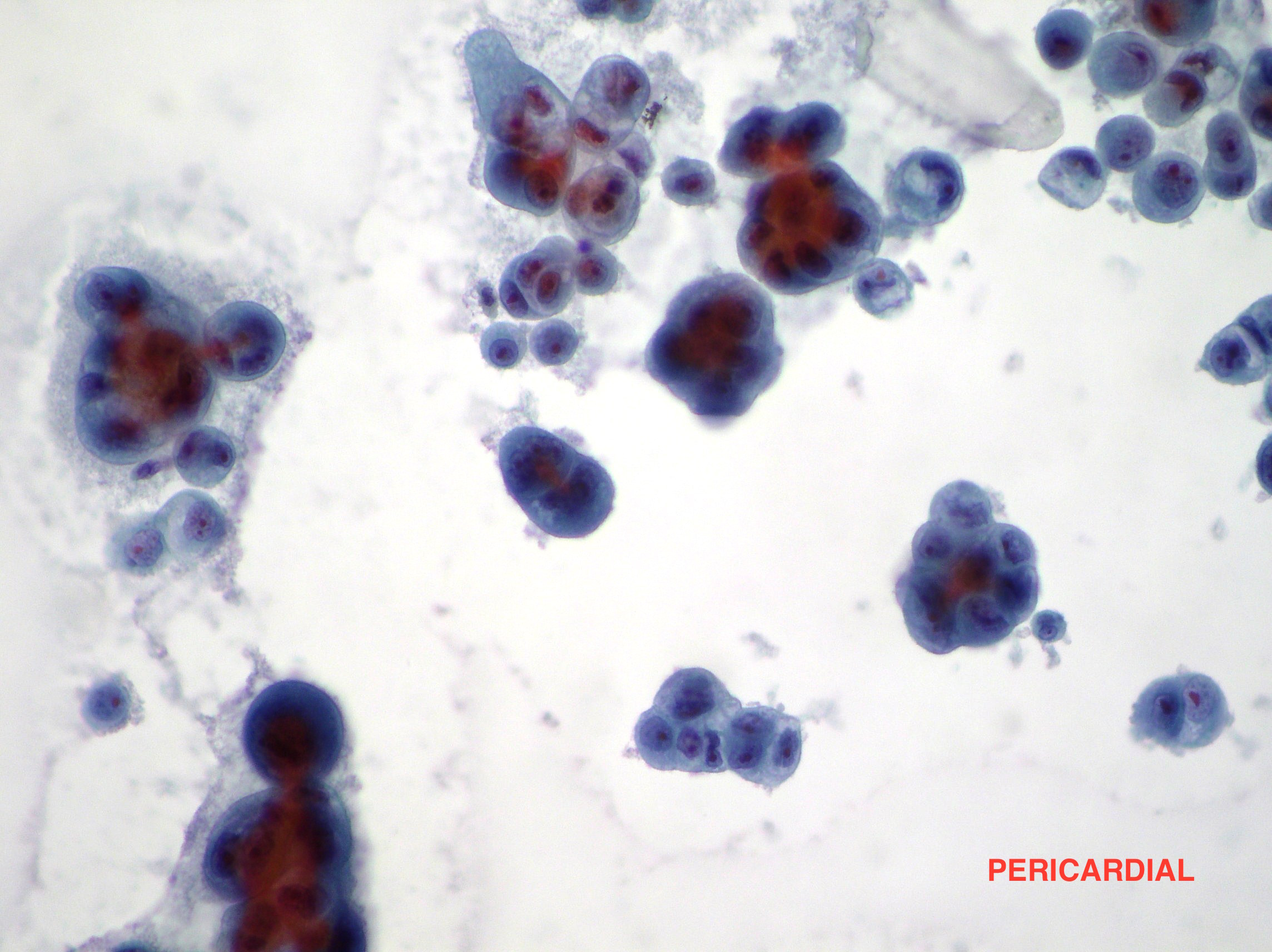
Adenocarcinoma cells in pericardial effusion
Lung adenocarcinoma cells (TTF-1+; CK-7+) observed in a case of pericardial malignant effusion. (CellBlock, Papanicolaou x200)
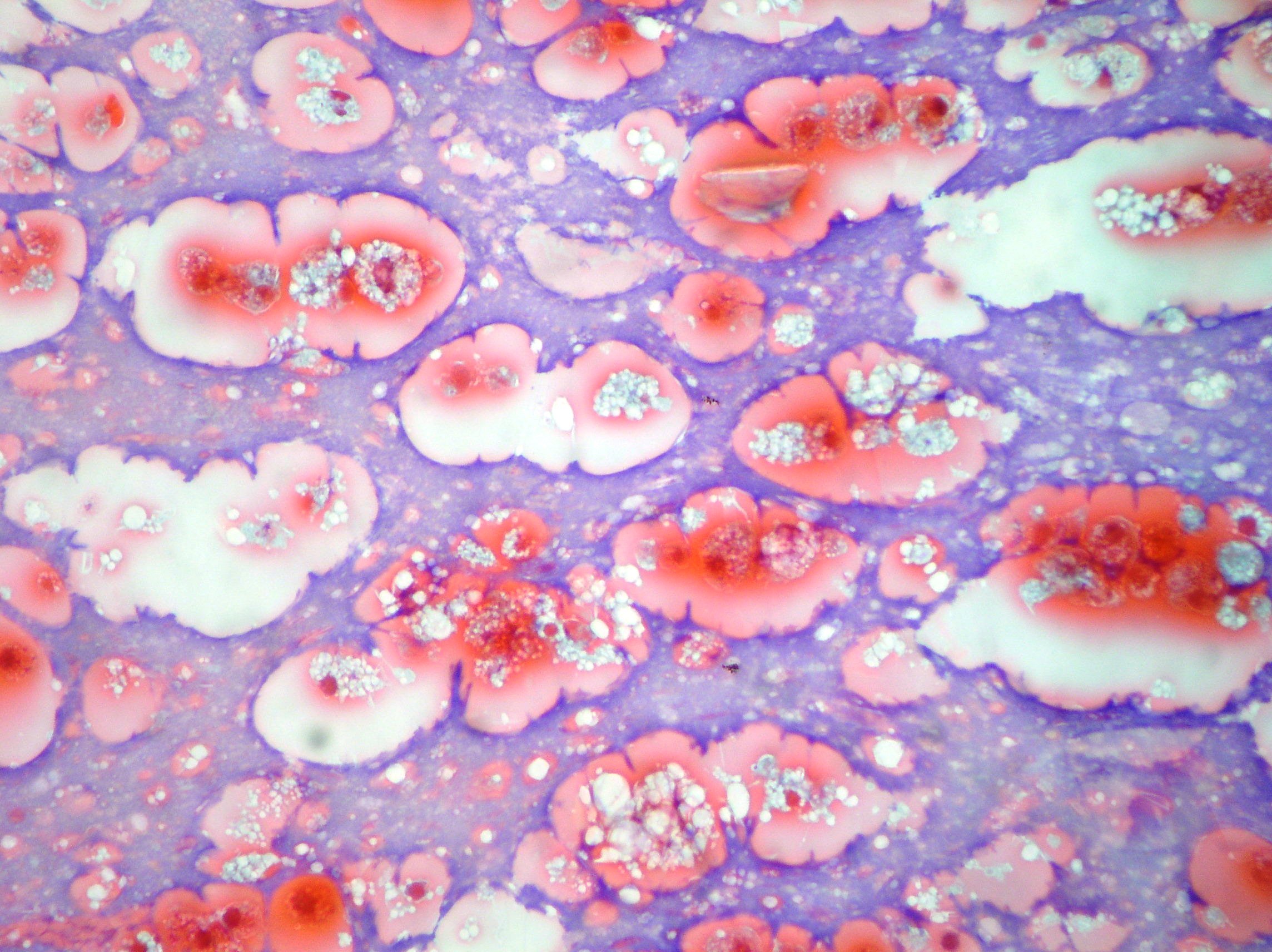
Pleural effusion
Defects of sample preservation in a case of pleural mesothelioma. Mesothelial cells are observed with marked degenerative effects. (Papanicolaou x200)
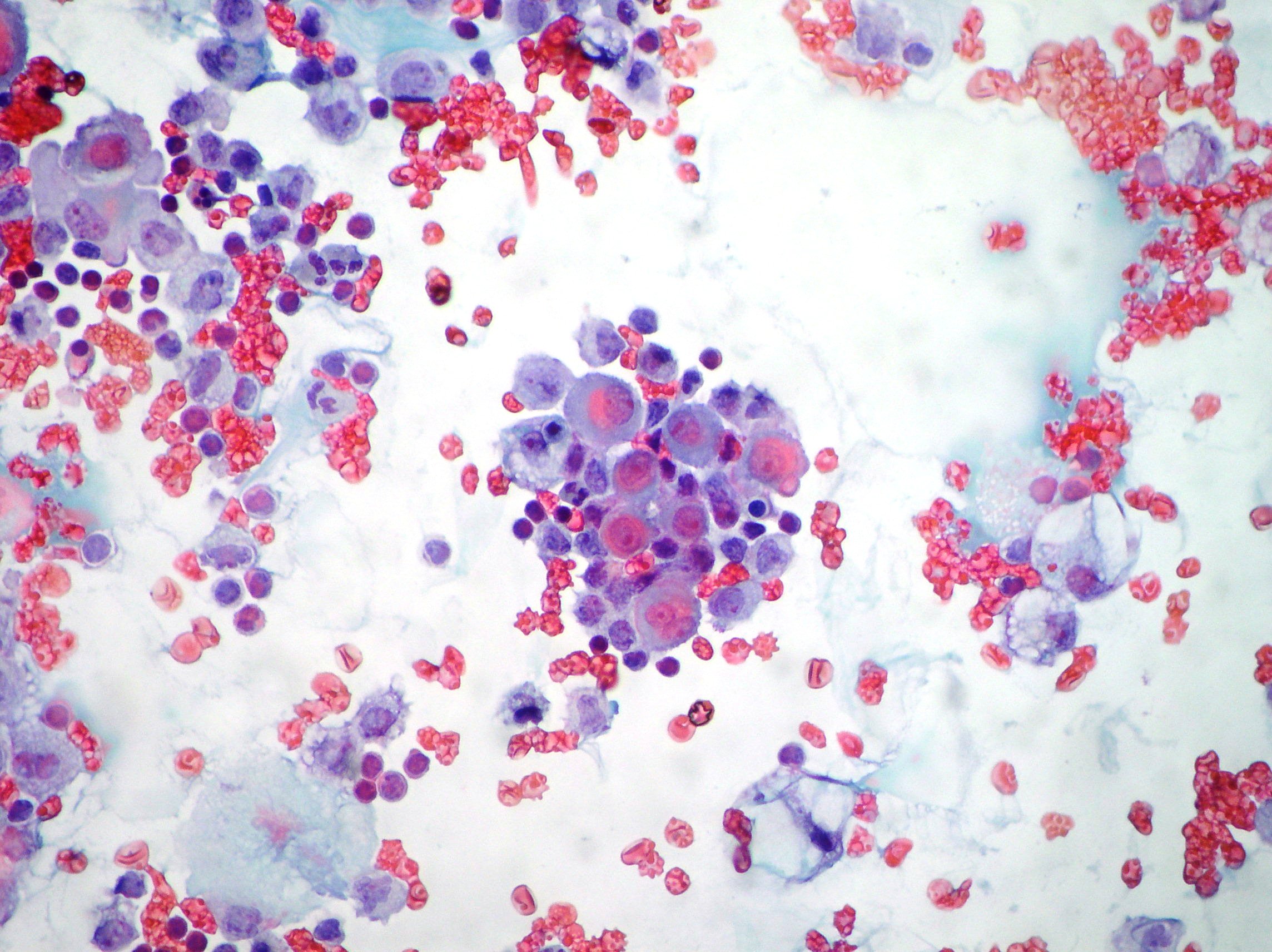
Pleural effusion
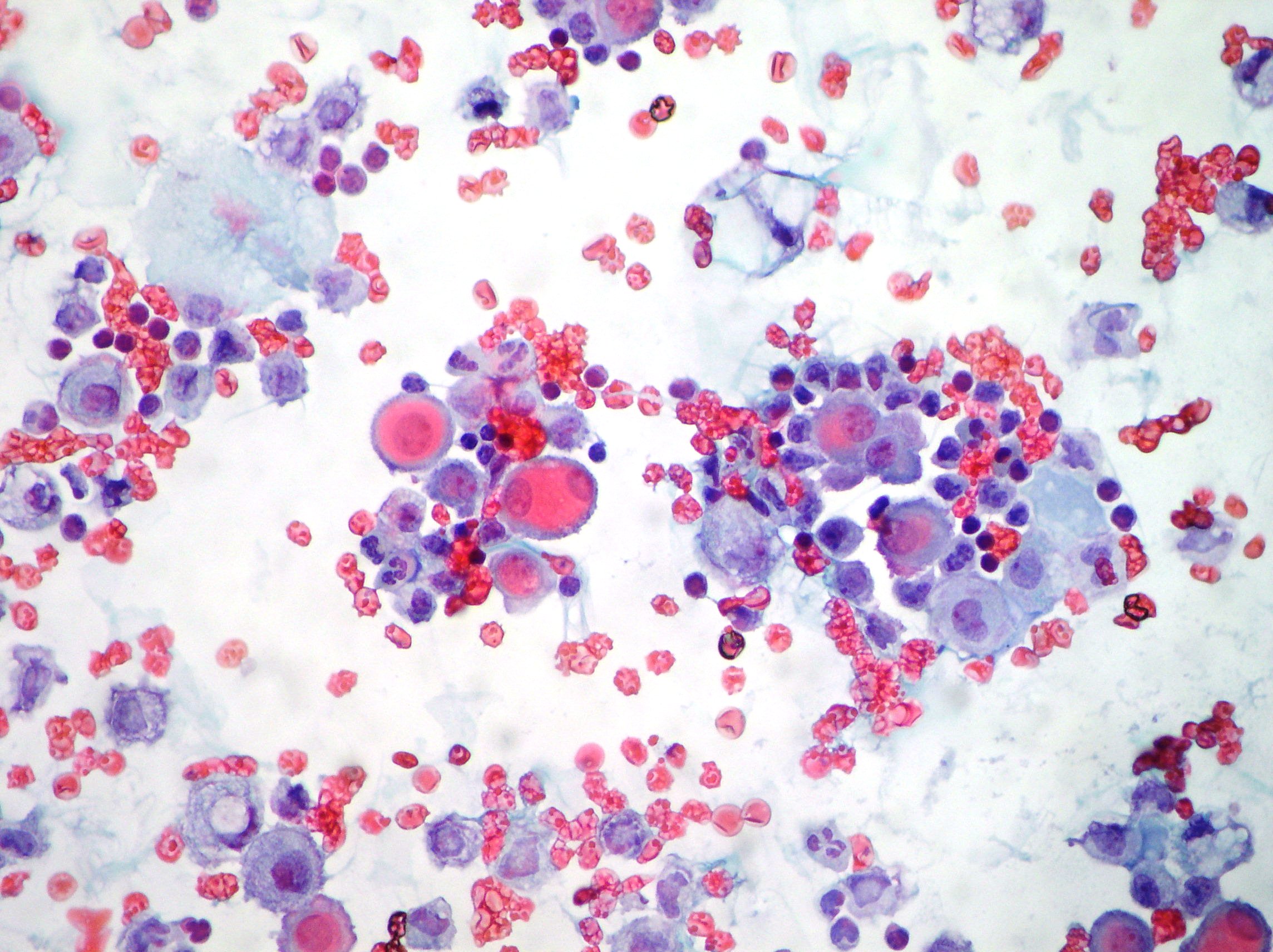
Reactive Pleural effusion showing mesothelial cells, lymphocytes, neutrophils and macrophages. (Papanicolaou x200)
Pleural effusion
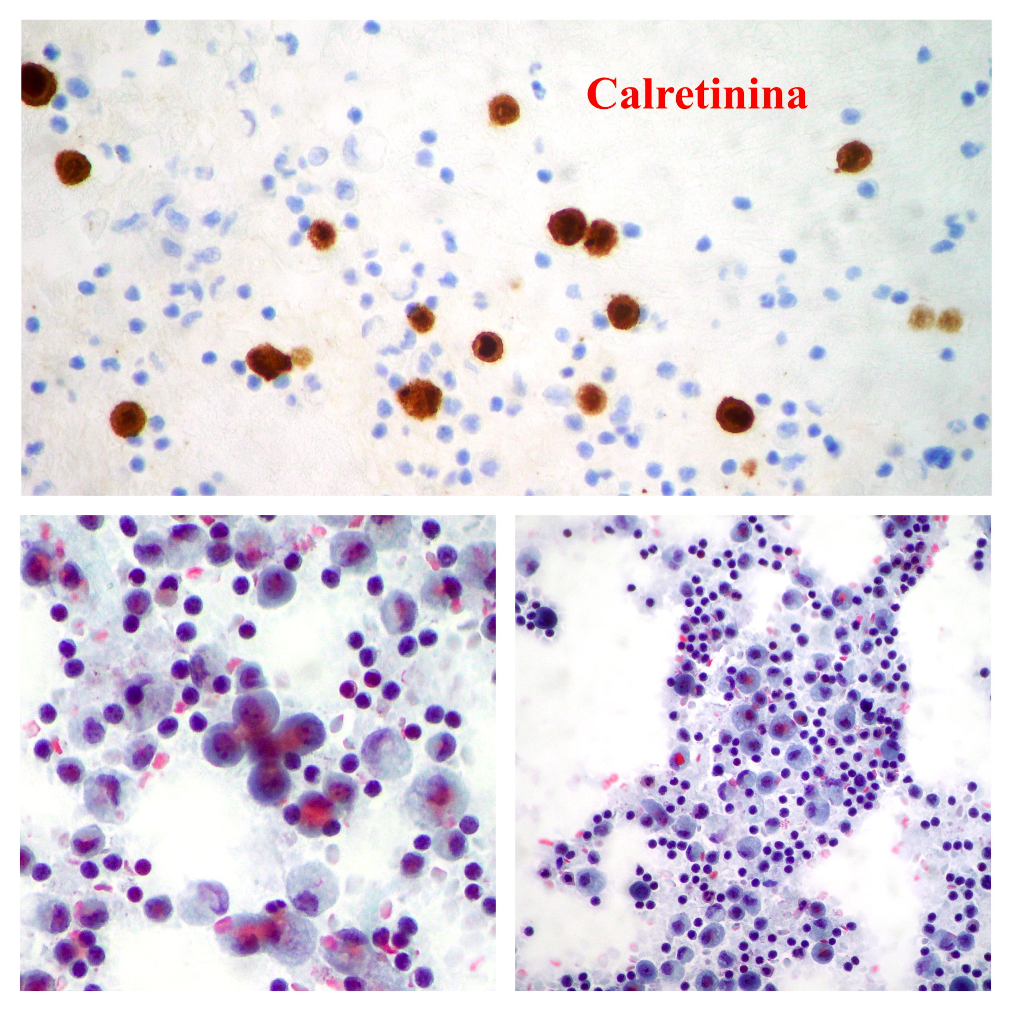
Reactive Pleural effusion showing mesothelial cells, lymphocytes, neutrophils and macrophages. Immunocytochemistry test resulted positive for Calretinin marker antibody (Papanicolaou x100, x200)
Pleuric Mesothelioma
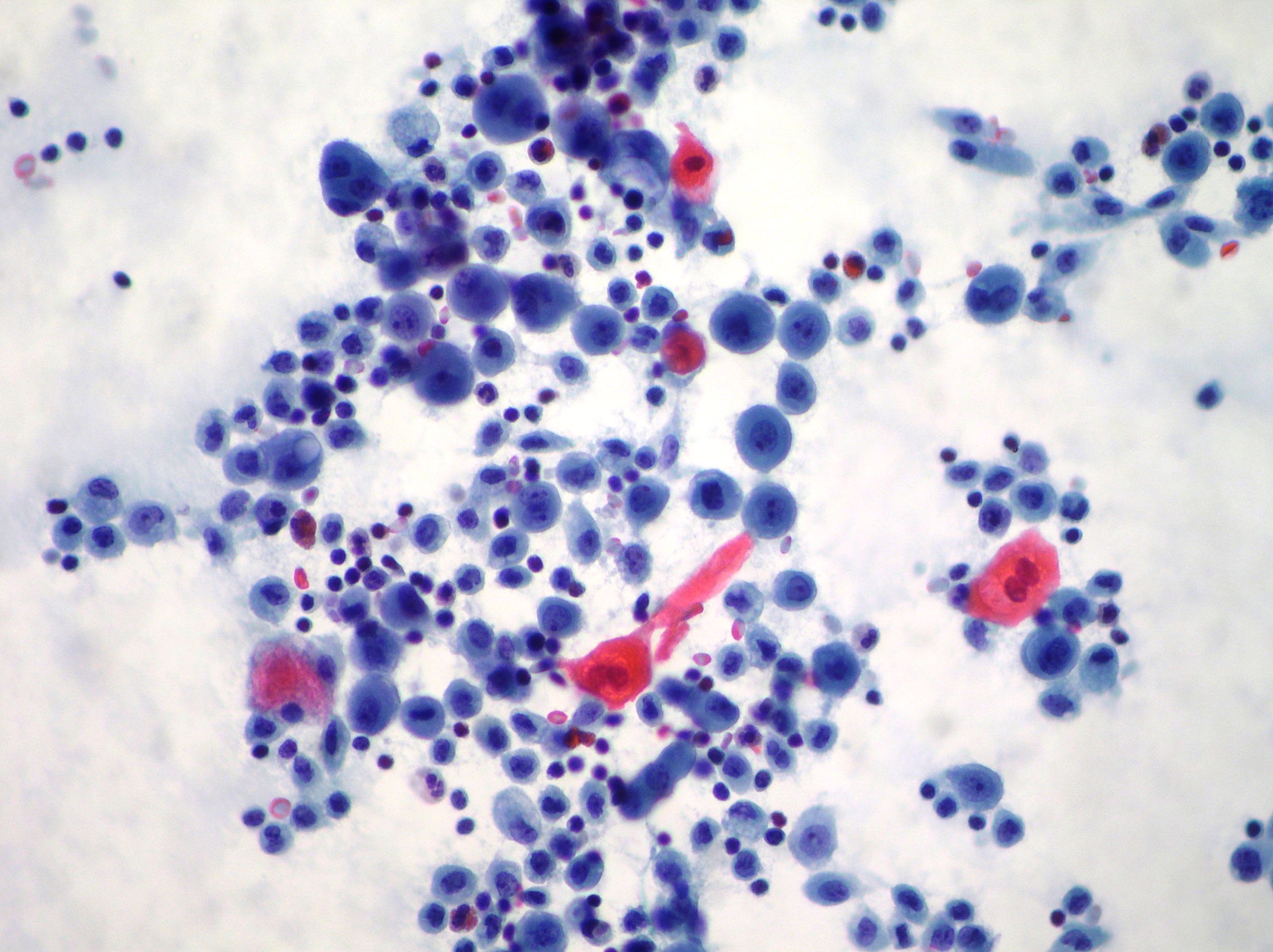
Pleural effusion showing isolated neoplastic cells some of theme with eosinophilic staining from a case of epithelioid mesothelioma.(Papanicolaou x100)
Adenocarcinoma in pleural effusion
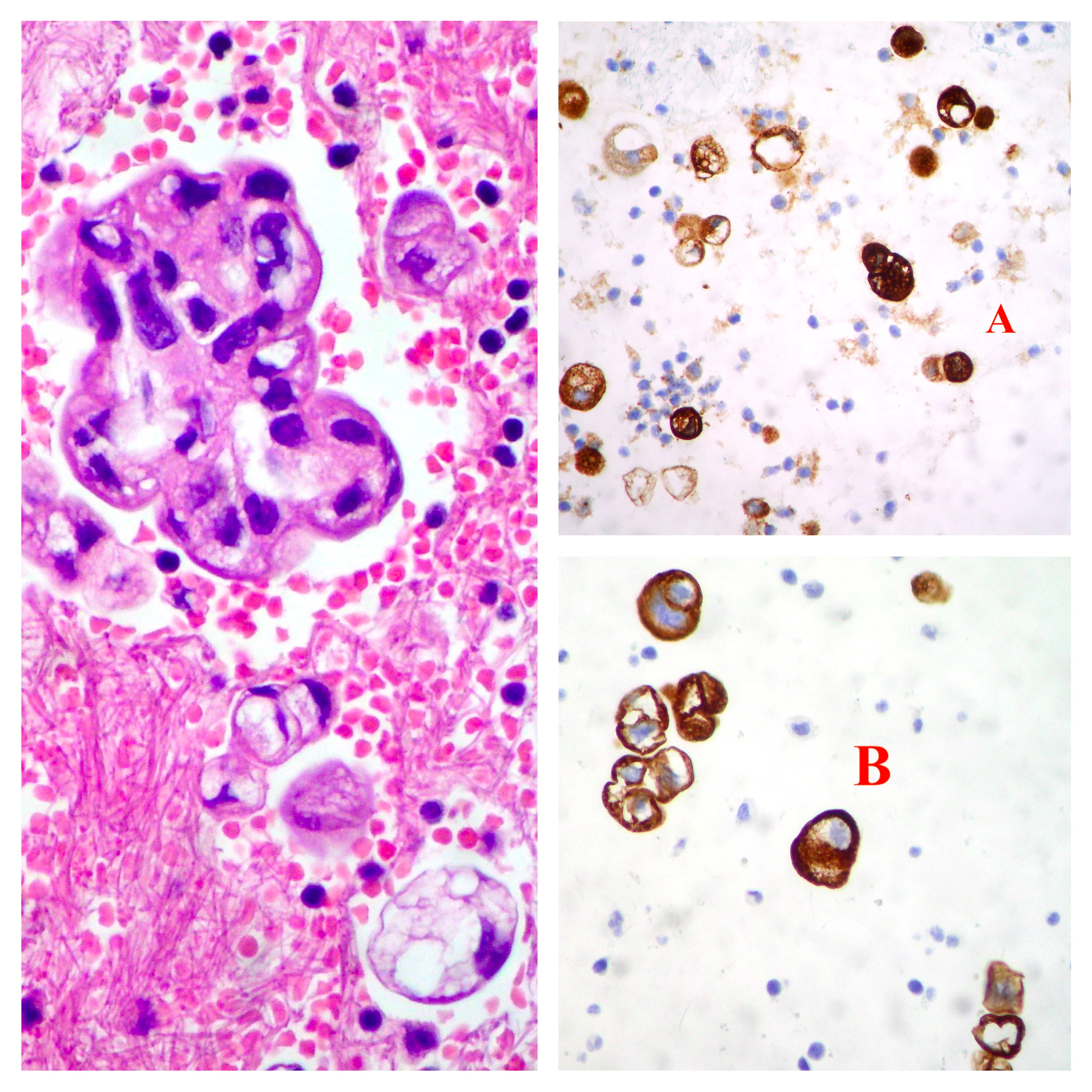
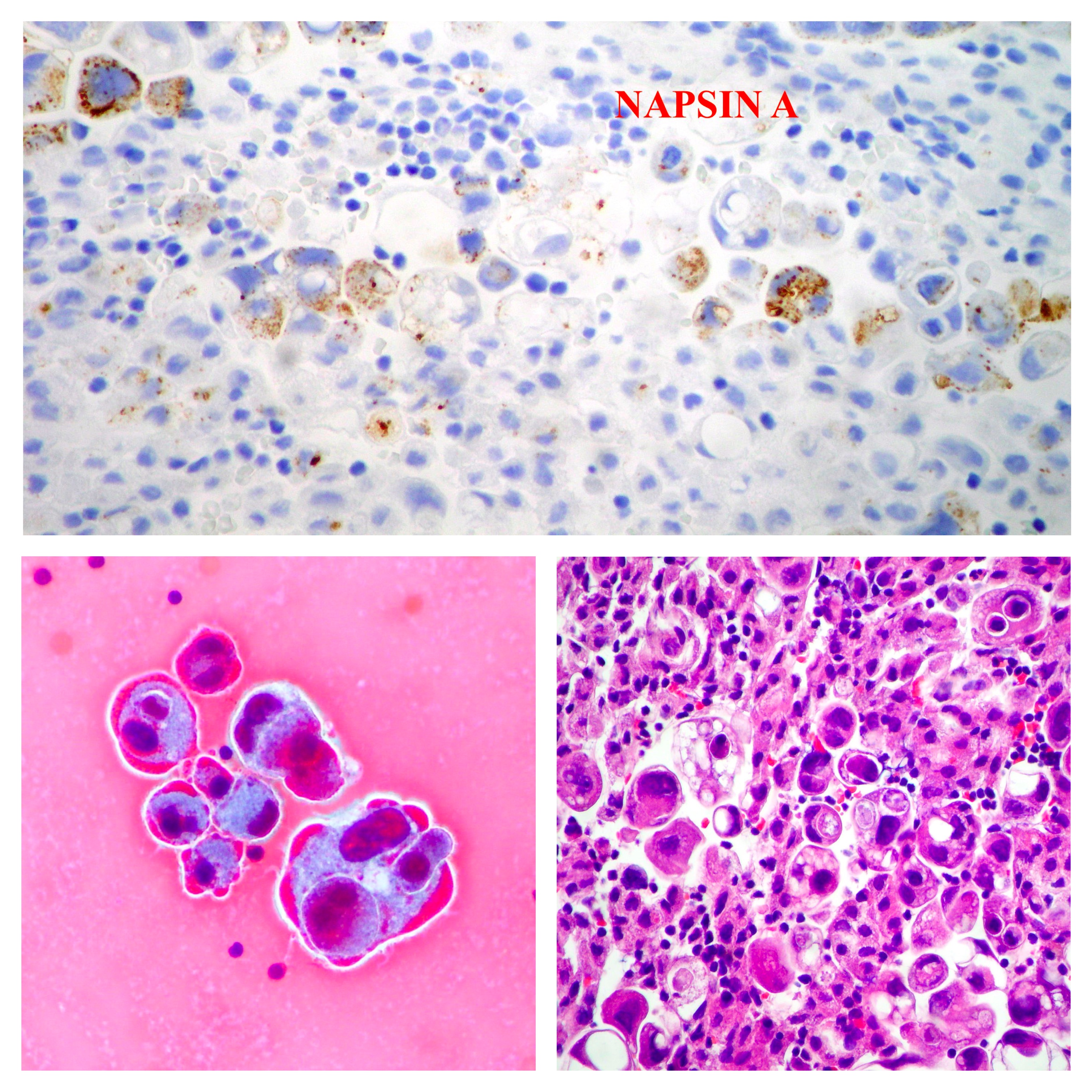
Pleural effusion showing isolated and clusters of neoplastic cells with reactivity for Napsin A by immunocytochemistry. (Cellblok H&E, Papanicolaou x200)
Pancreatic cancer cells in pleural effusion
Pleural effusion showing isolated and clusters of neoplastic cells with reactivity for CEA (A) and CK19 (B) by immunocytochemistry. (Cellblok Hematoxylin Eosin x200)
Neuroendocrine pancreatic cancer cells in abdominal effusion
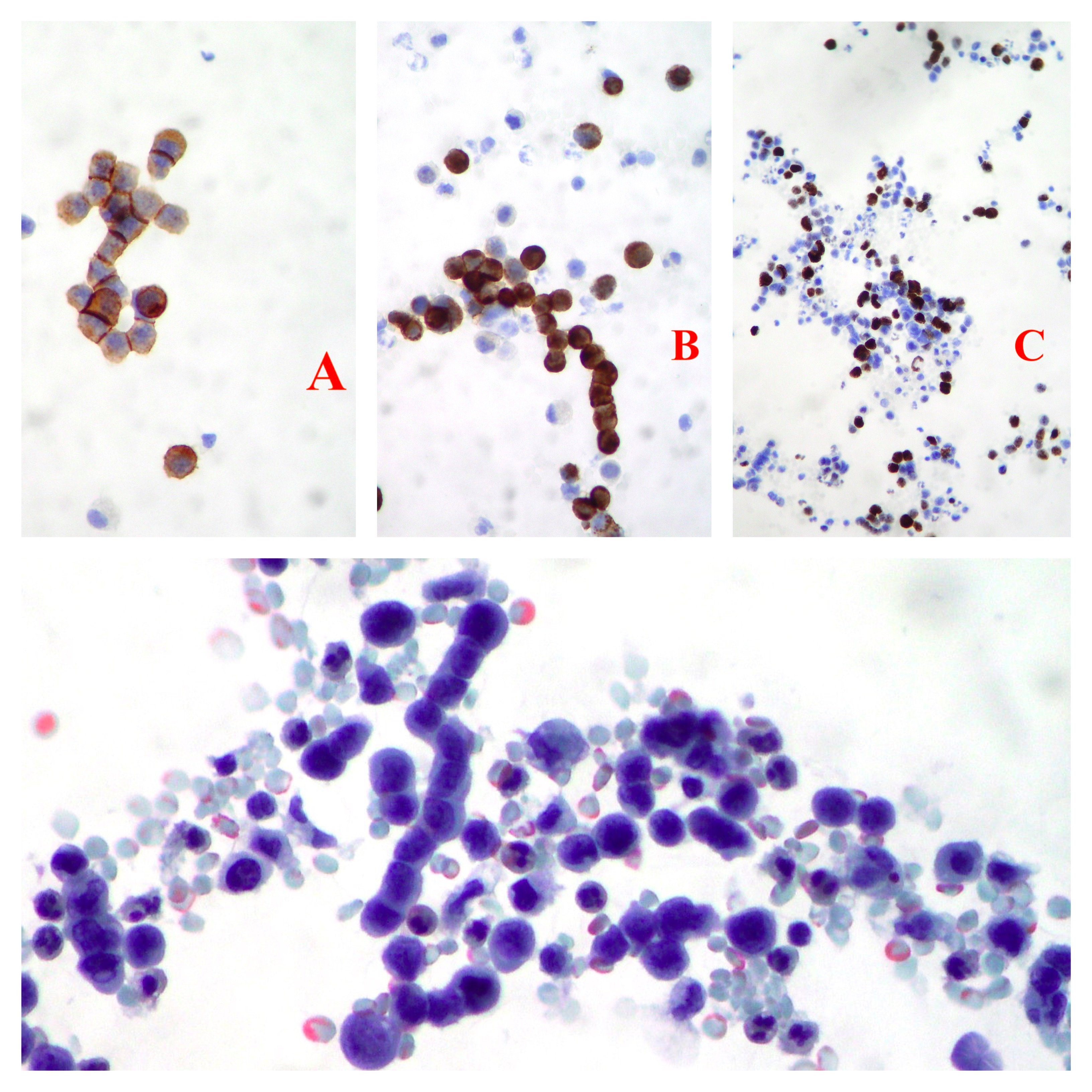
Abdominal effusion showing isolated, groups and chains with molding of neuroendocrine neoplastic cells. Positivity for CD56 (A), Chromogranin (B) and Ki67 (C) by immunocytochemistry is observed. (Papanicolaou x200)
Ovary cancer cells in pleural effusion
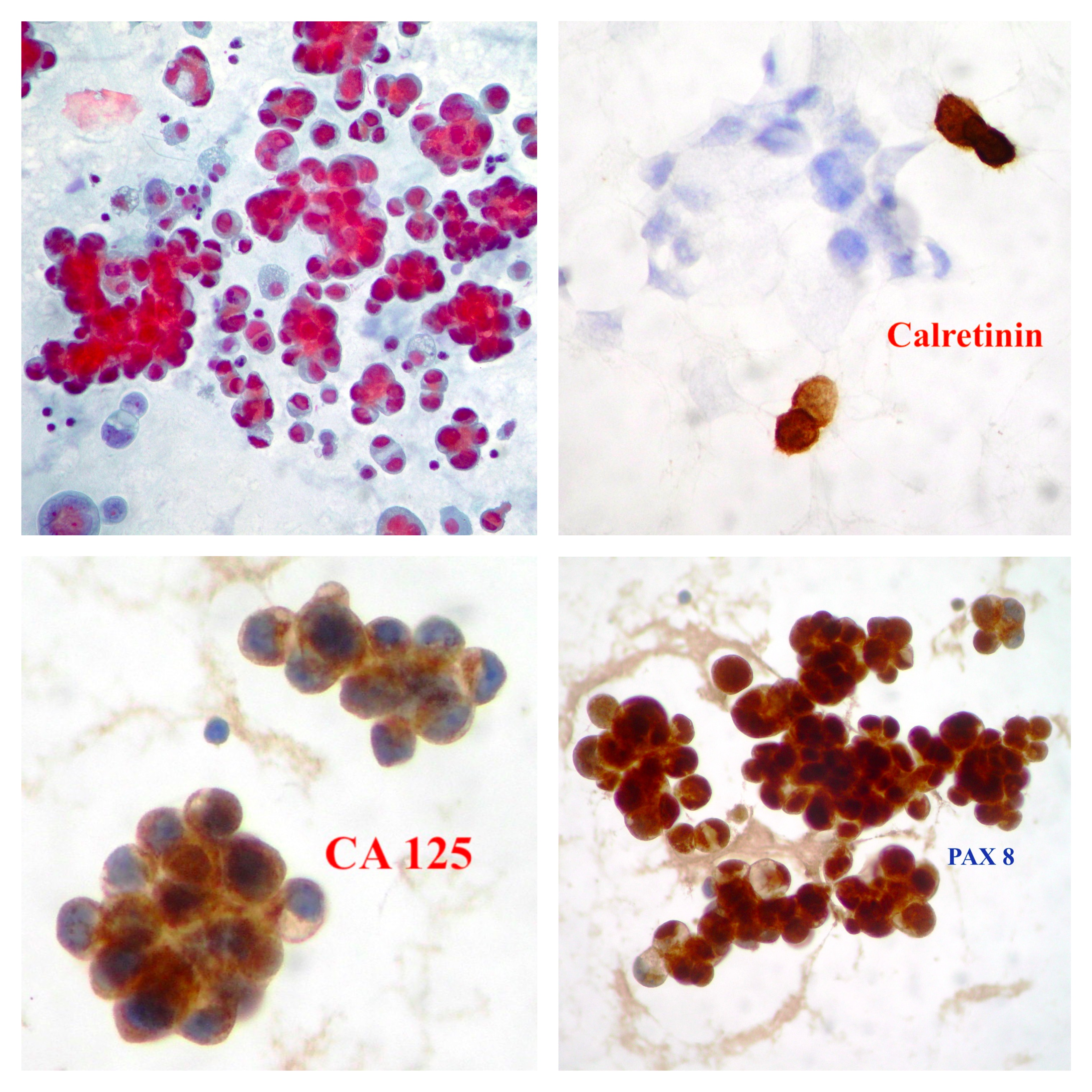
Pleural effusion showing clusters of neoplastic cells from a case of metastatic serous papillary ovary cancer CA125 (+), PAX 8 (+) and Calretinin (-) by immunocytochemistry. (Papanicolaou x200)
Breast cancer cells in pleural effusion
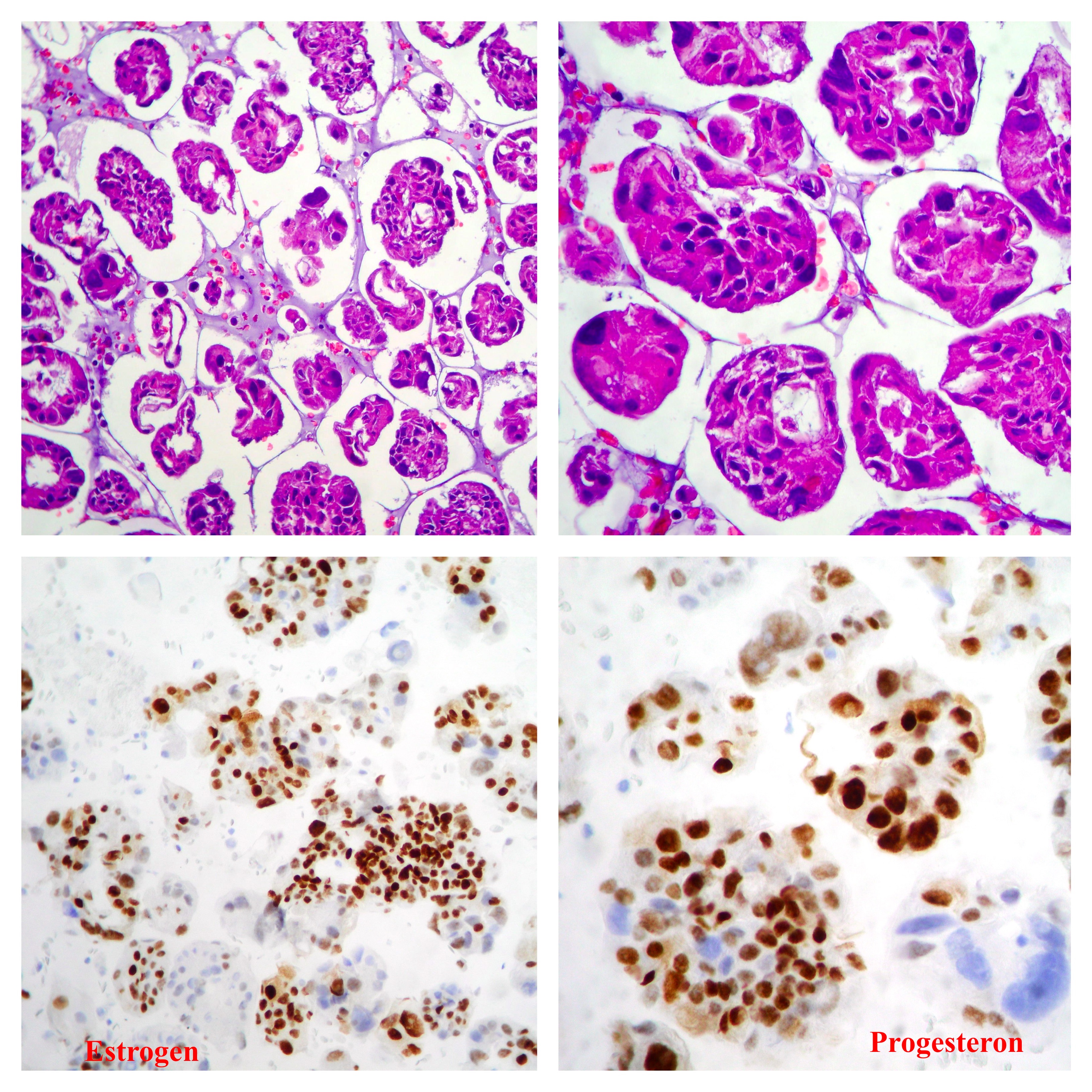
Cell block. Pleural effusion showing clusters of neoplastic cells from a case of metastatic breast cancer Er and PgR positive by immunohistochemistry.(Hematoxylin Eosin x100, x200)
Pleuric Mesothelioma
Pleural effusion showing a cluster of neoplastic cells from a case of malignant mesothelioma.(Papanicolaou x100 oil immersion)
Ovarian cancer cells in abdominal effusion
Isolated and pseudopapillary groups of neoplastic cells associated to psammoma bodies and inflammatory cells. CA125 (on the left) and Napsin A (right) resulted positive by immunocytochemistry. (Papanicolaou x400, x200)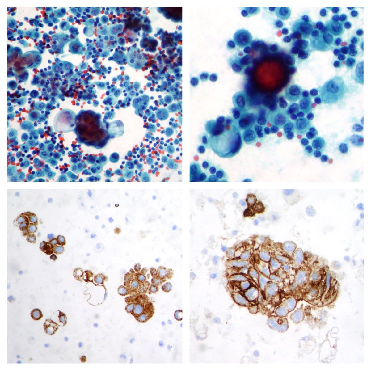
Malignant pleural effusion cells
Adenocarcinoma cell clusters / balls with large citoplasmatic vacuolization (Papanicolaou, x200)
Lung adenocarcinoma cells in pleural effusion
Lung adenocarcinoma cells “mimicking” mesothelial cells. The immunocytochemistry resulted TTF-1 positive. (Papanicolaou x100, x200) 
Ovarian cancer cells in abdominal effusion
A case of ovarian cancer cells CA125 positive by immunocytochemistry. (Papanicolaou x400, x200) 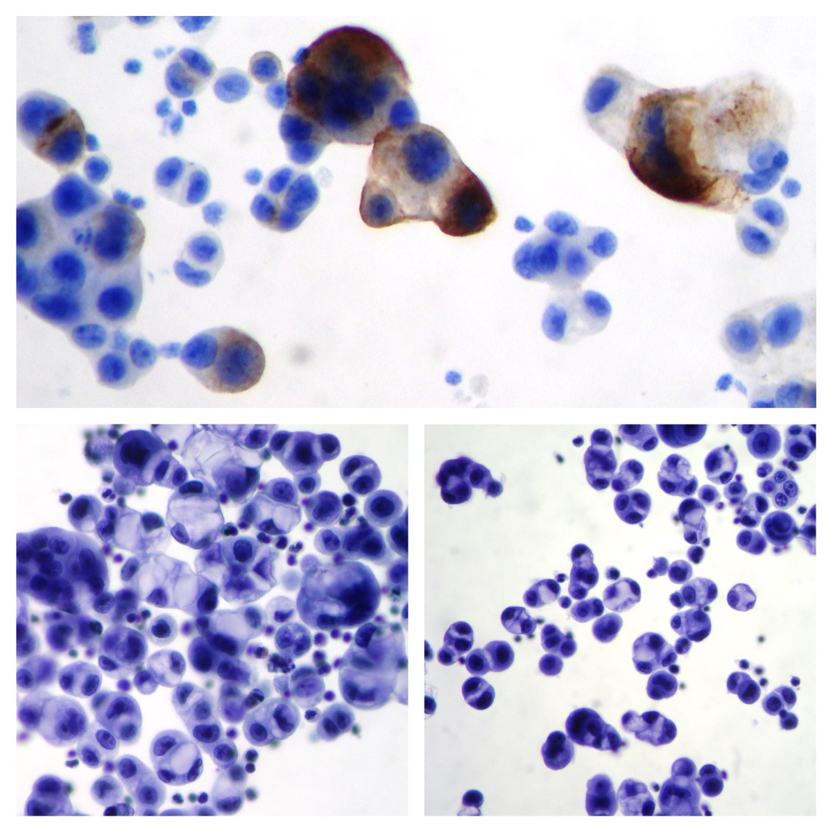
Pancreatic cancer cells in abdominal effusion
A case of pancreatic cancer. Morphology and immunocytochemistry support the origin of the cells. (Papanicolaou x 200)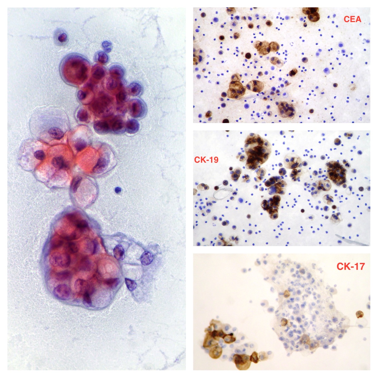
Mesothelial cells
Reactive pleural effusion. Normal mesothelial cells. (Papanicolaou x200)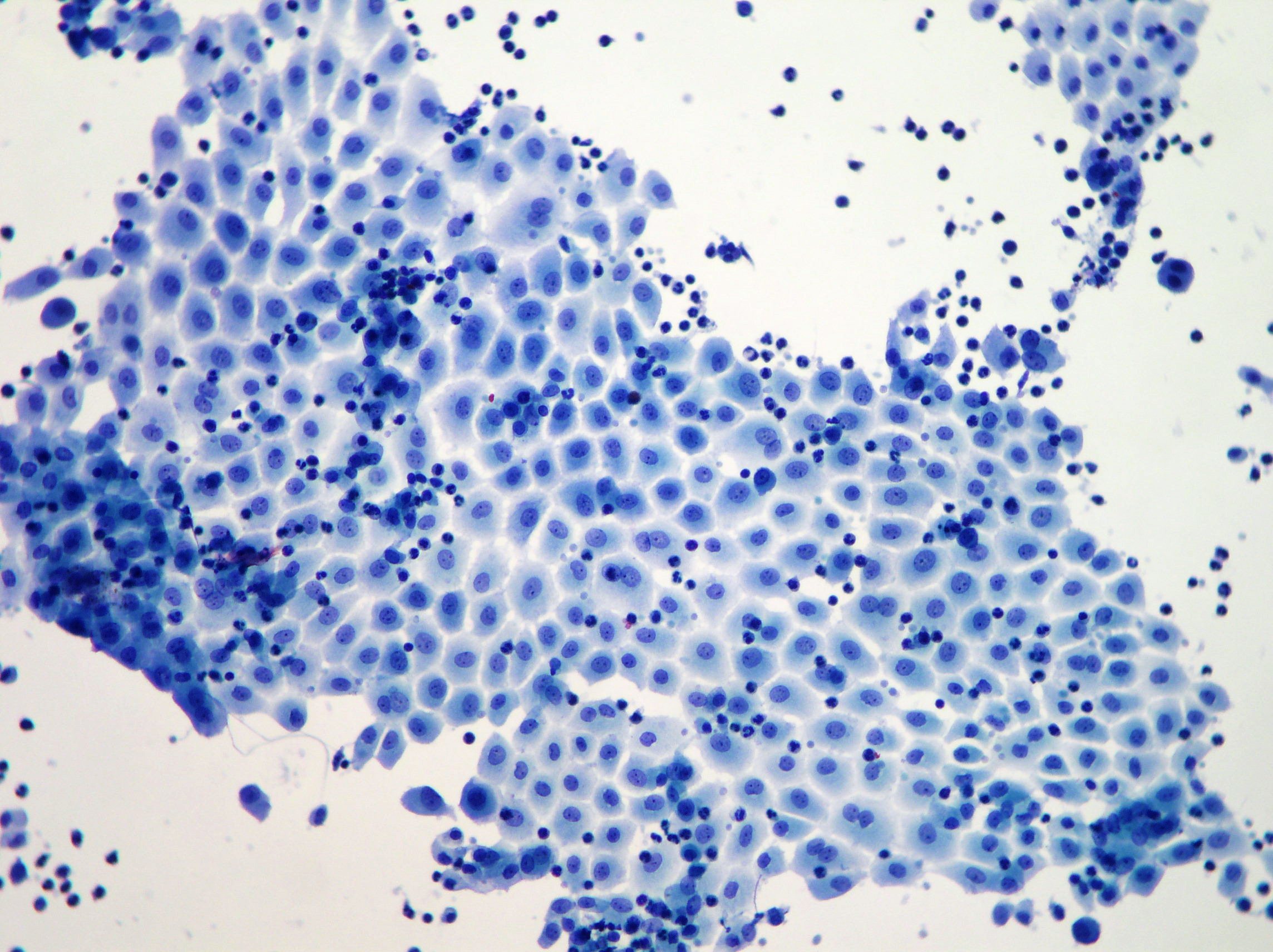
Breast carcinoma cells in pleural effusion
Metastatic lobular breast carcinoma Estrogen Receptor positive by immunocytochemistry. (Papanicolaou x200)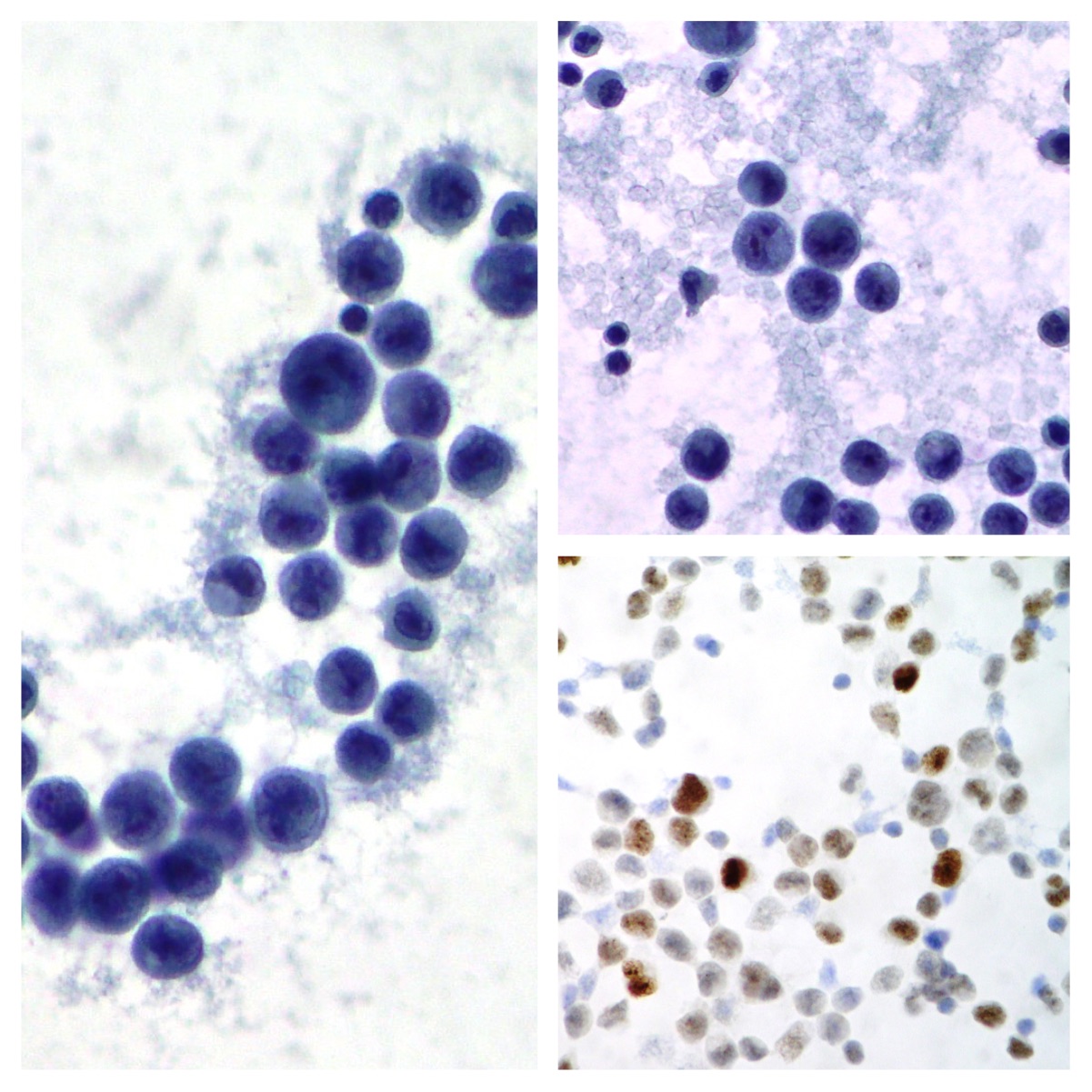
Adenocarcinoma cells in pleural and pericardial effusion
Lung adenocarcinoma cells observed in a case with concomitant pleural and pericardial malignant effusion (Papanicolaou x200)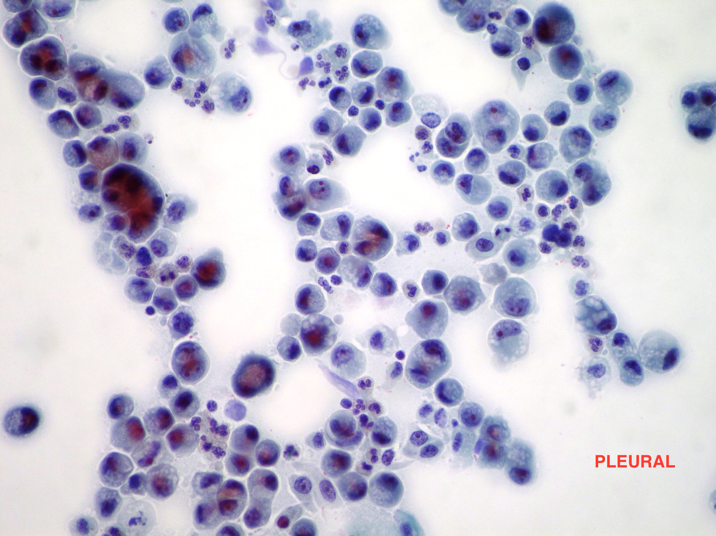
Lung adenocarcinoma cells in pleural effusion
Malignant pleural effusion cells from lung adenocarcinoma . Immunoreactivity for CK 7 (left) and TTF-1 (right) confirmed the diagnosis. (Papanicolaou x100, x200)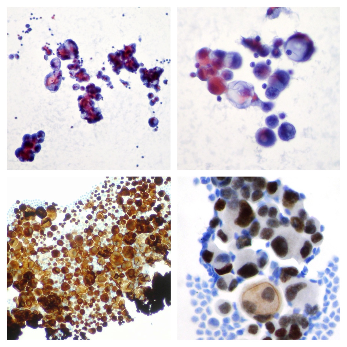
Ovarian adenocarcinoma cells in abdominal effusion
Peritoneal effusion. Cells of mucinous ovarian adenocarcinoma (Papanicolaou x100, x400)
Squamous cell carcinoma in pleural effusion
Metastatic squamous cell carcinoma of the lung (Papanicolaou, x200)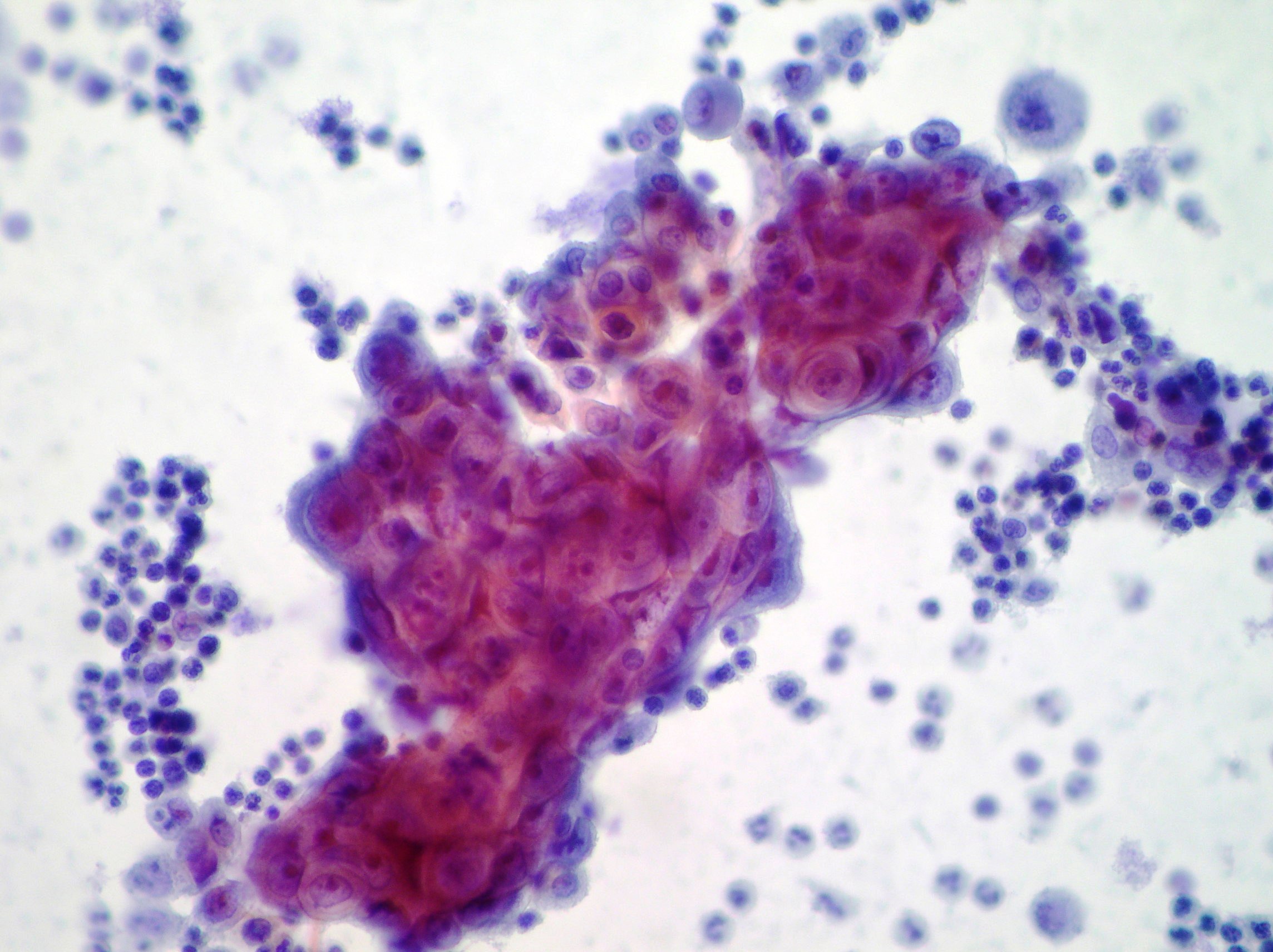
Adenocarcinoma cells in pleural effusion
Adenocarcinoma cells (Papanicolaou, x200)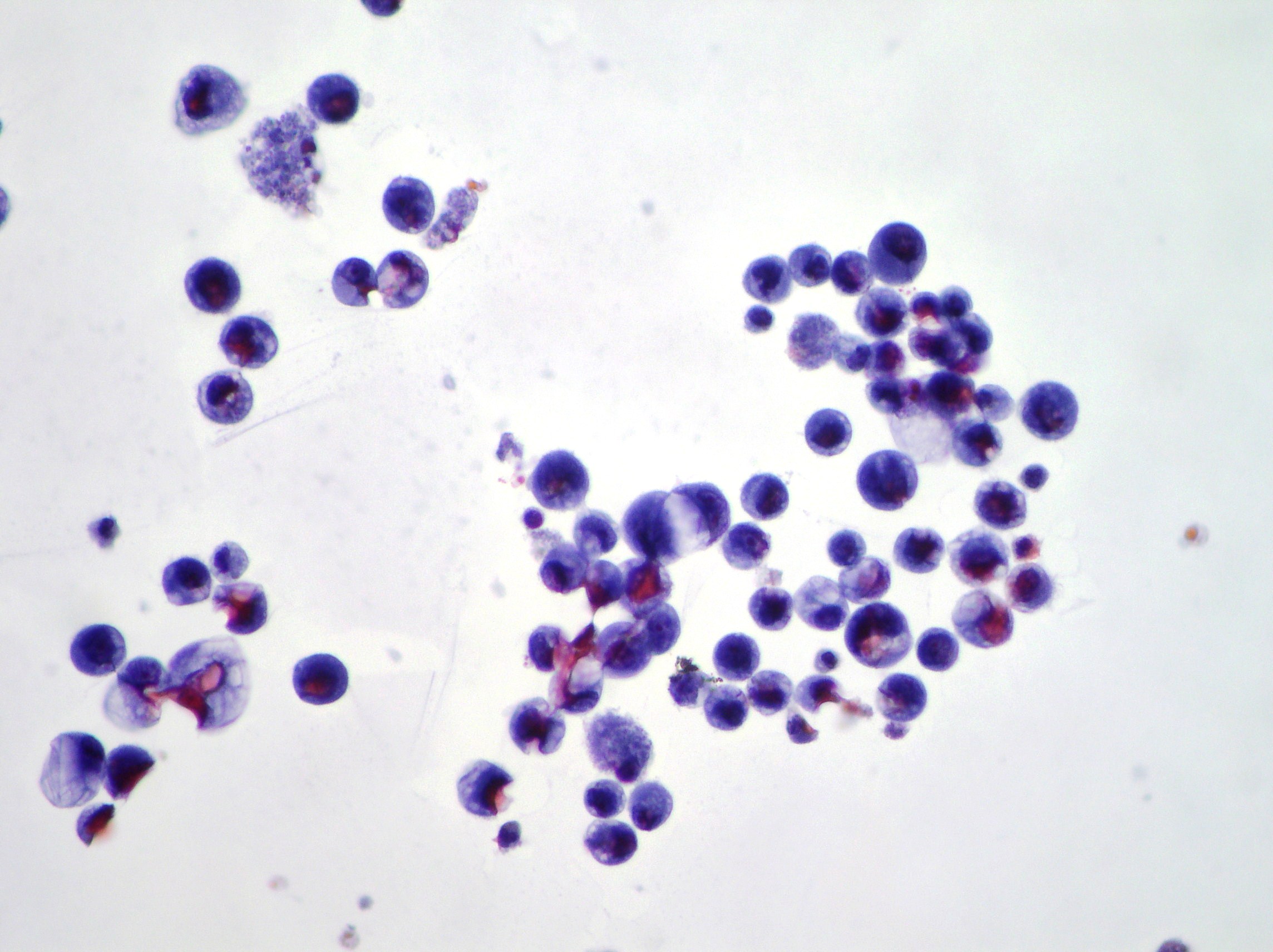
Normal mesothelial cells
Pleural effusion showing monolayered sheet of normal mesothelial cells demonstrating “spongiotic” separation of individual cells within the sheet. (Papanicolaou, x100)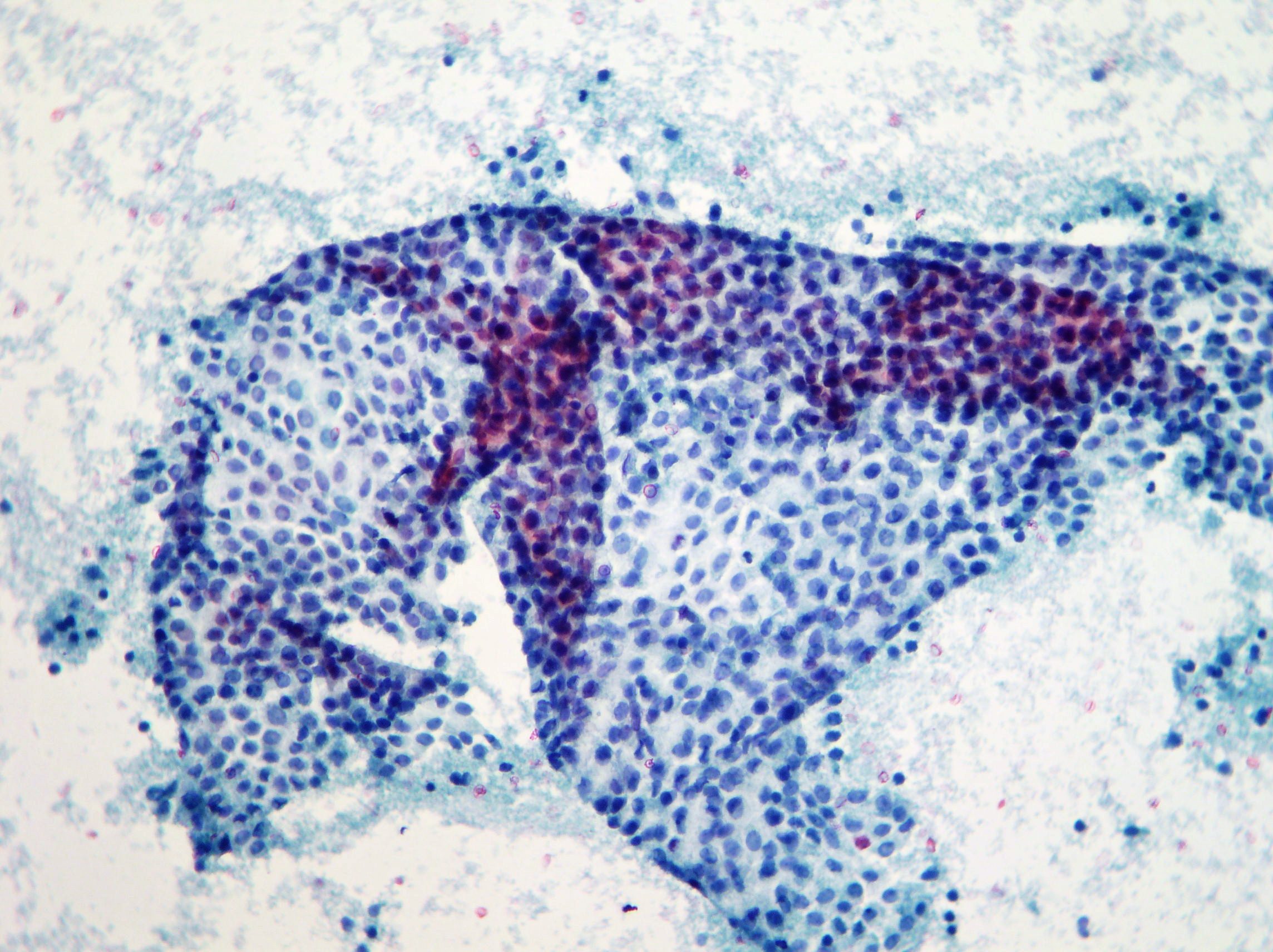
Reactive pleural effusion
Reactive pleural effusion showing acute and chronic cells, normal mesothelial cells and alveolar macrophages in aggregates and dispersed cells with rounded nuclei and vacuolated cytoplasm. (Papanicolaou, x100)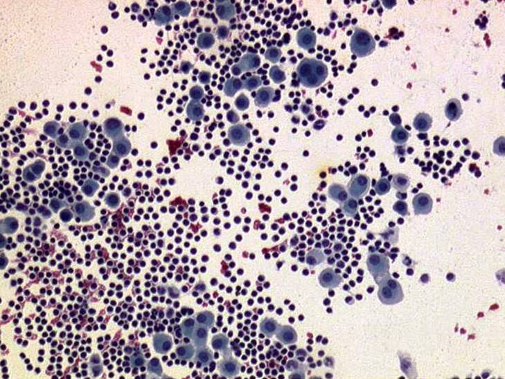
Breast adenocarcinoma cells in pleural effusion.
Spheroid aggregate of metastatic ductal cell adenocarcinoma of the breast in a smear of pleural effusion. (Papanicolaou, x100) 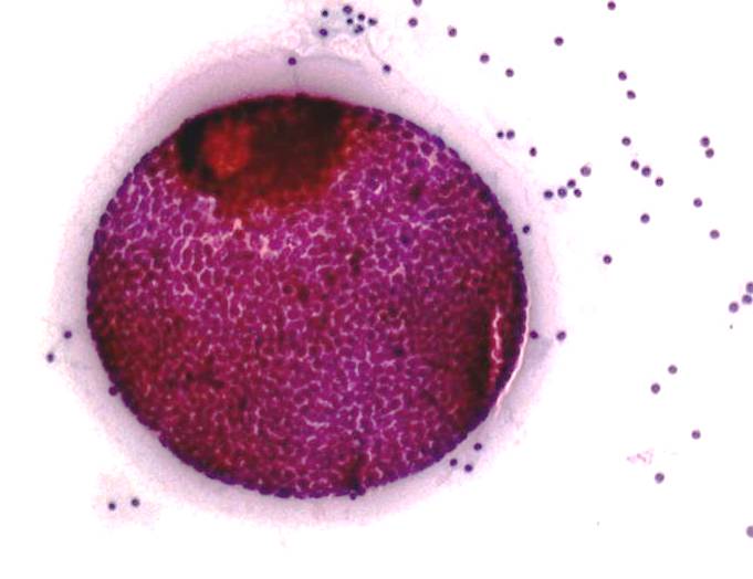
Lung adenocarcinoma cells in pleural effusion
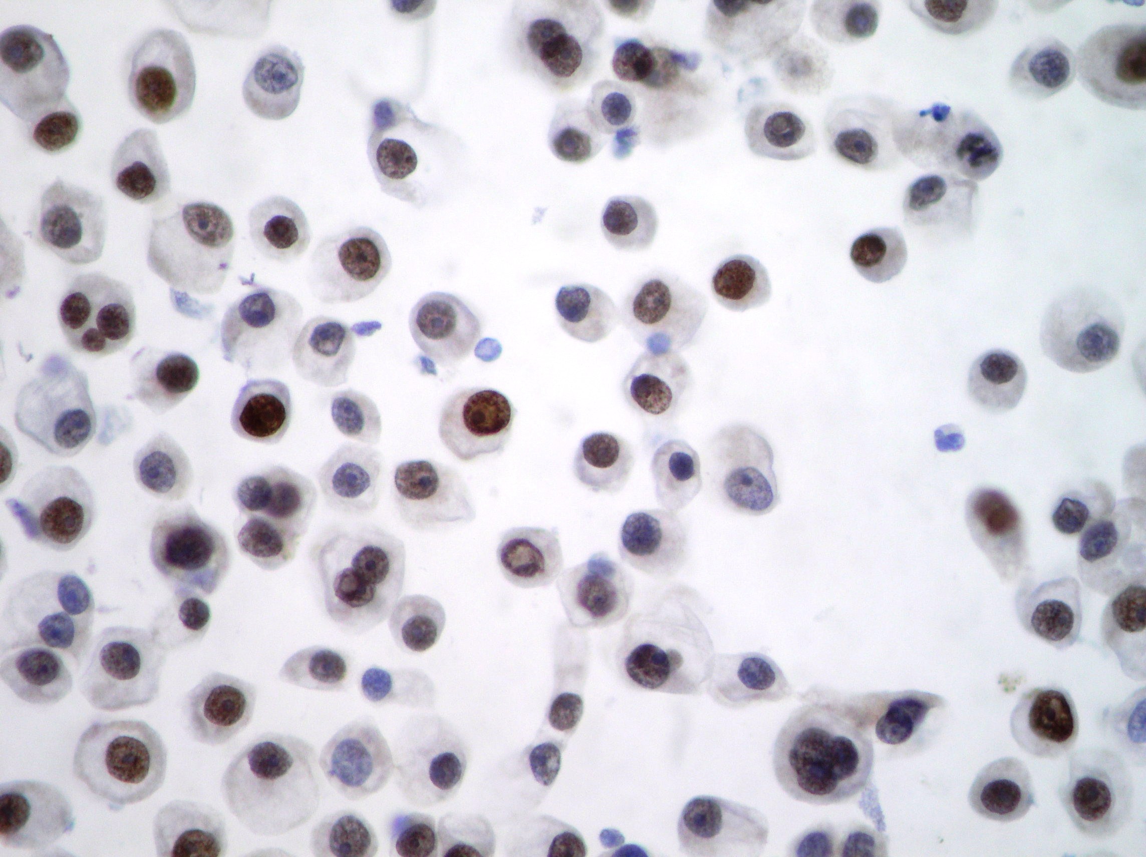
Lung adenocarcinoma pleural effusion showing pseudopapillary clusters of cancer cells. TTF-1 immunocytochemistry shows nuclear positivity confirming lung origin of the cells. (Papaniclaou, x200)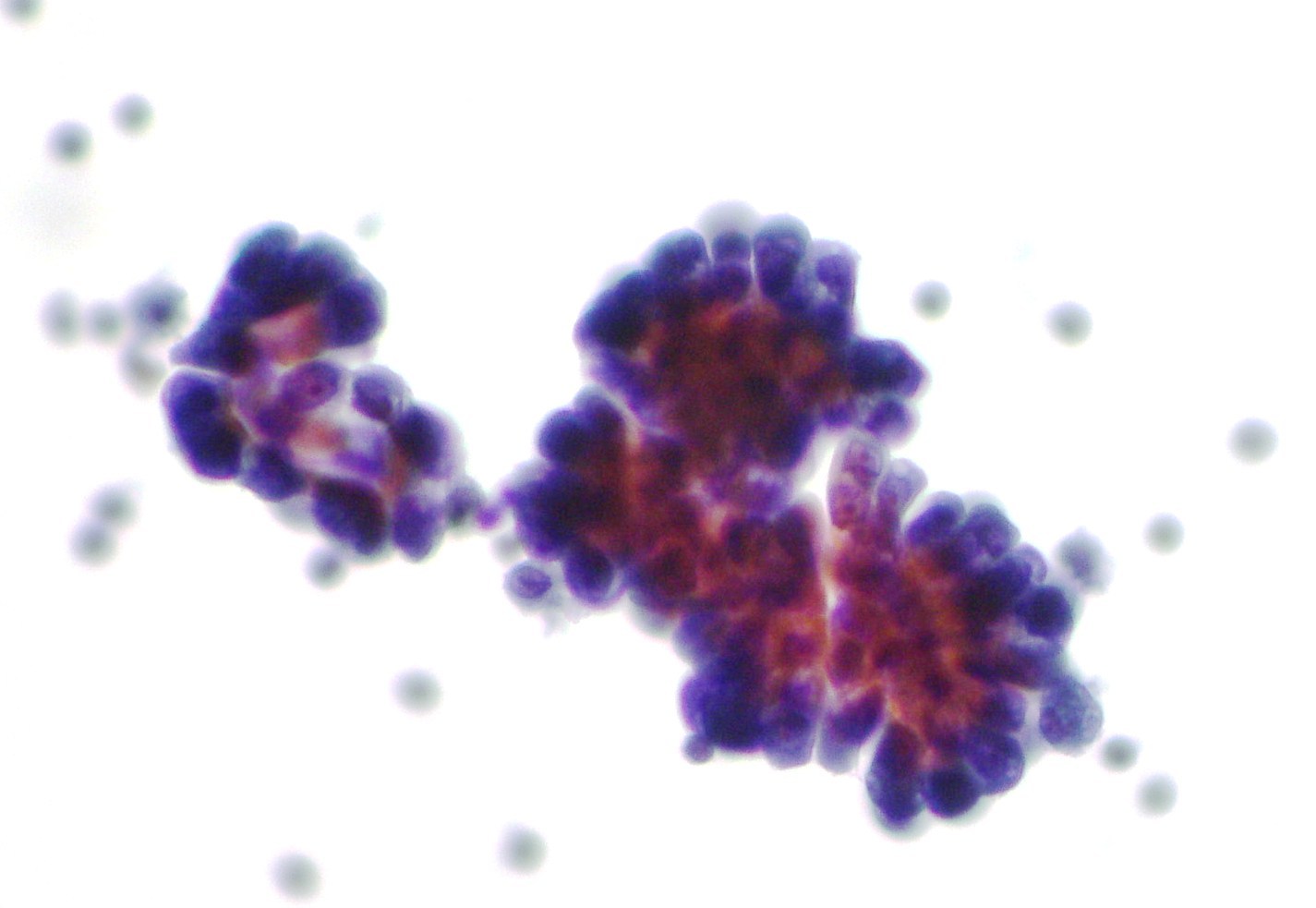
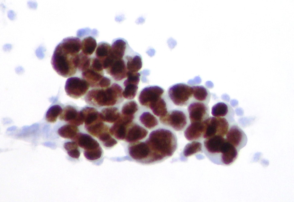
Ovarian adenocarcinoma cells in abdominal effusion
Smear of peritoneal effusion showing cells of metastatic ovarian papillary serous adenocarcinoma containing a psammoma body (on the left). (Papanicolaou, x200)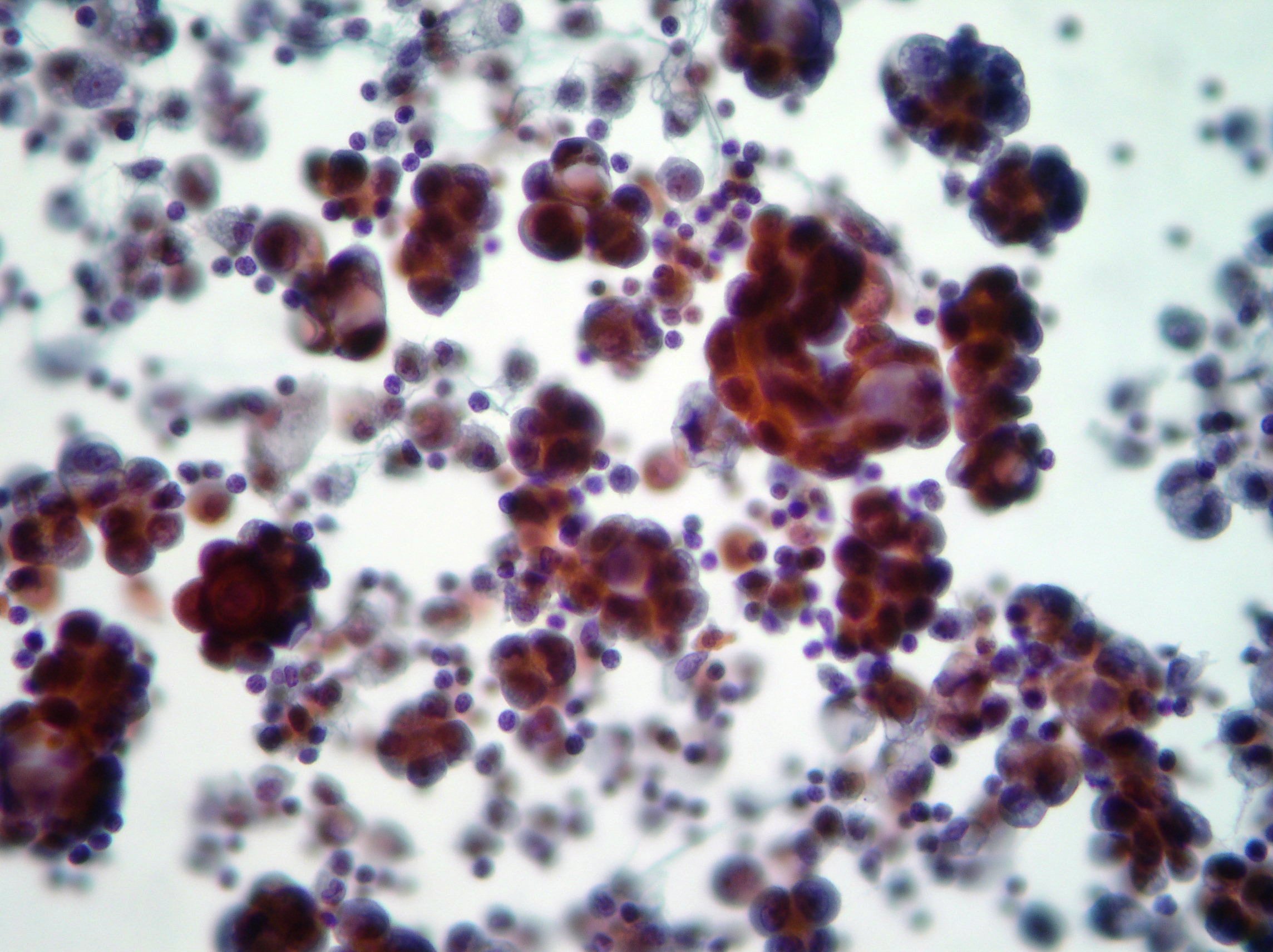
Mesothelioma pleural effusion.
Smear of pleural effusion showing papillary fragments of malignant mesothelioma. (Papanicolaou, x200, x400) 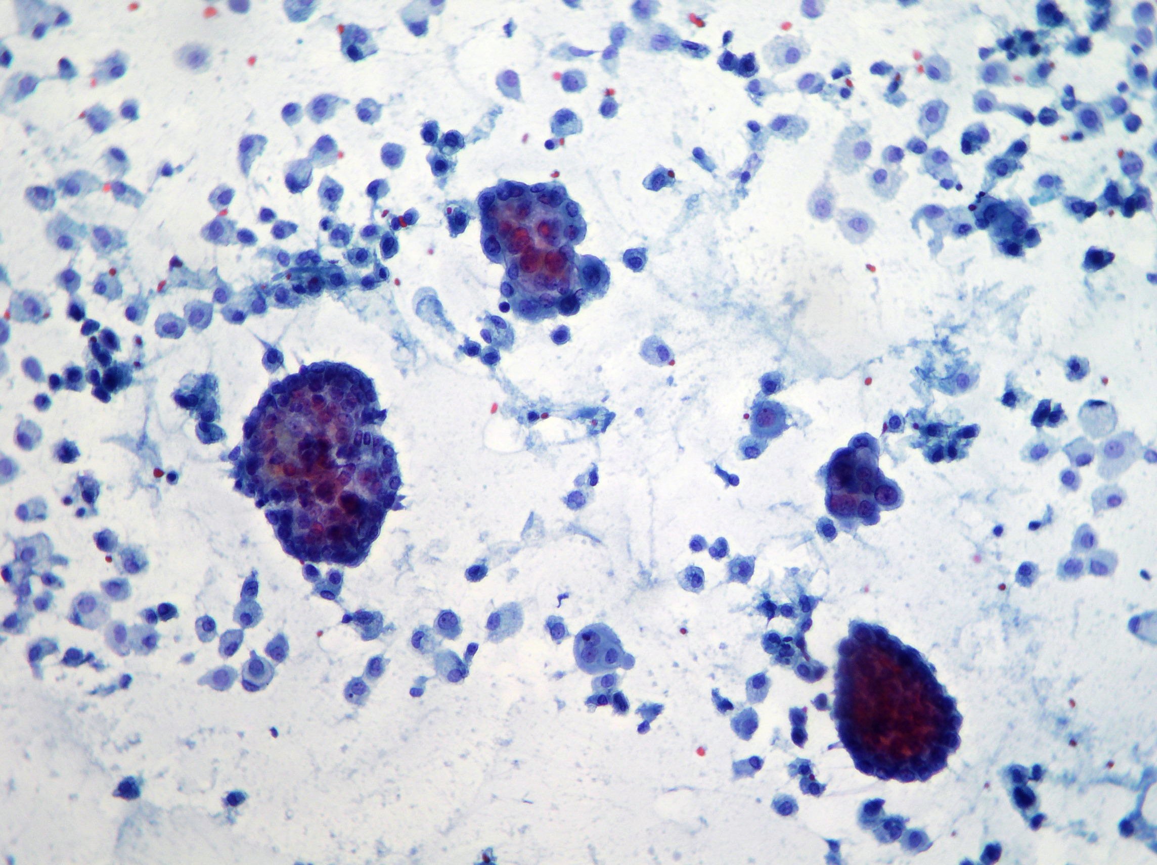
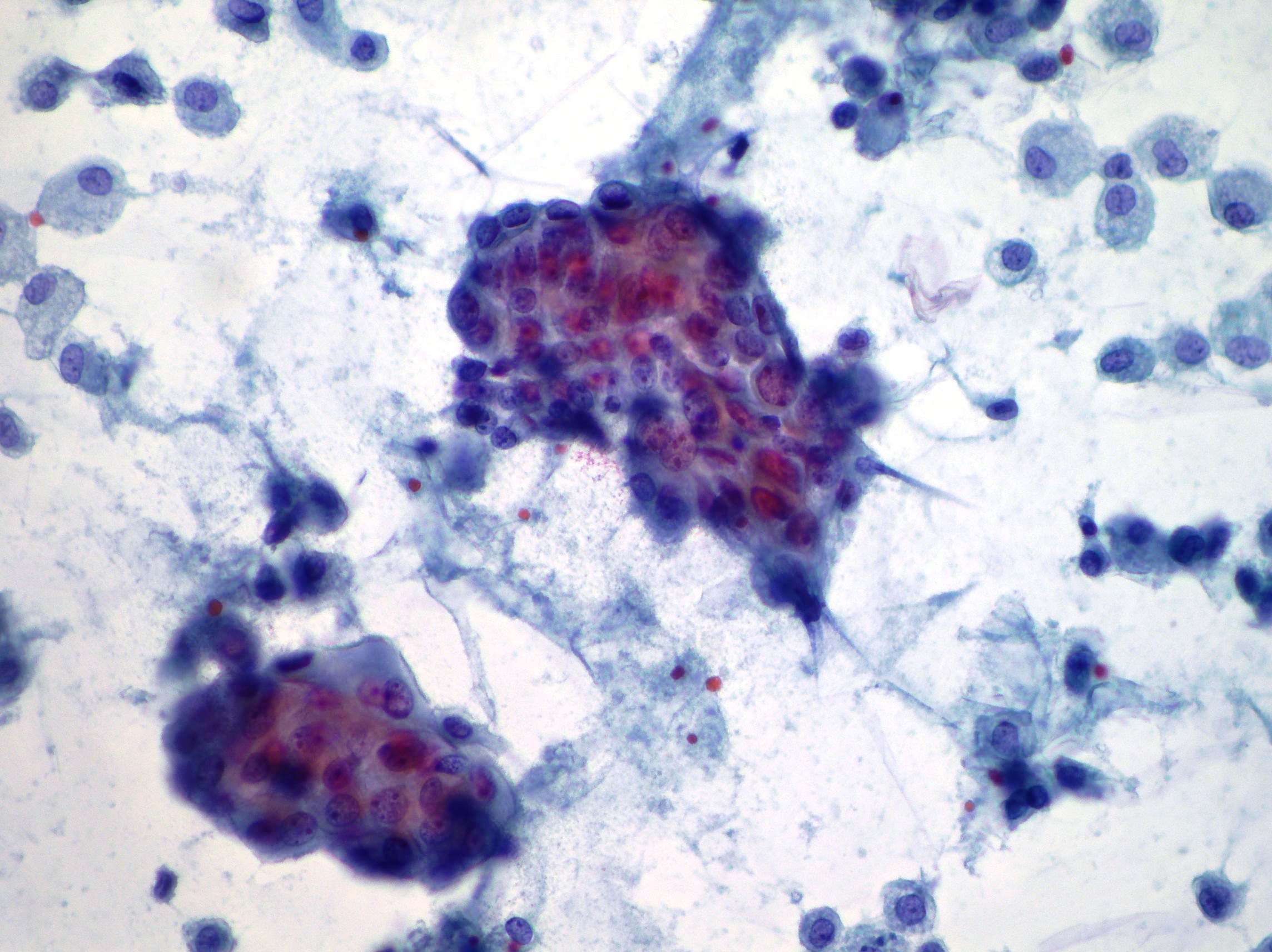
Small cell carcinoma cells in pleural effusion
Smear of pleural effusion showing small cell carcinoma with typical tiny chain and molding. (Papanicolaou, x400)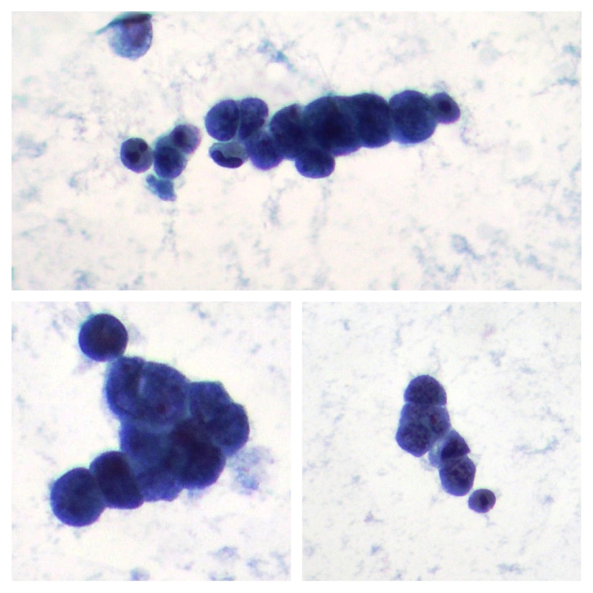
Ovarian adenocarcinomacells in abdominal effusion
Smear of peritoneal effusion showing metastatic ovarian adenocarcinoma cells resulted CA-125 positive. (Papanicolaou, x400)